
















Copyright: ©Author(s) 2026.
World J Gastroenterol. Jul 14, 2026; 32(26): 114936
Published online Jul 14, 2026. doi: 10.3748/wjg.114936
Published online Jul 14, 2026. doi: 10.3748/wjg.114936
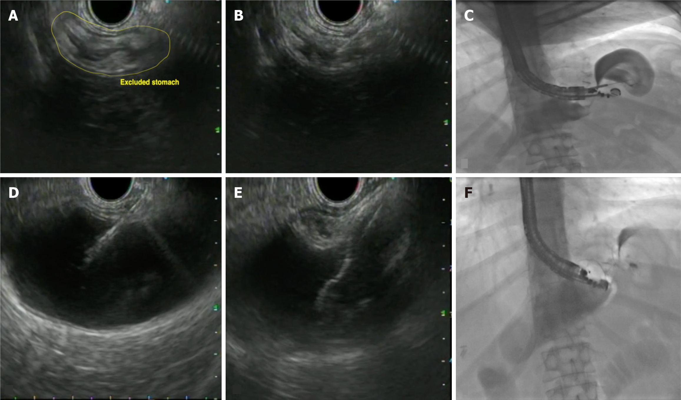
Figure 5 Endoscopic ultrasound-directed trans-gastric endoscopic retrograde cholangiopancreatography for management of biliary obstruction in altered anatomy.
A: Endoscopic ultrasound (EUS)-directed trans-gastric endoscopic retrograde cholangiopancreatography being performed in a patient for the management of choledocholithiasis in a Roux-en-Y gastric bypass anatomy; Using linear echoendoscope, under EUS and fluoroscopic guidance, the excluded stomach was located endosonographically from the remnant gastric pouch; B: Once an optimal position was confirmed, the remnant stomach was punctured using a 19-G EUS-fine needle aspiration (FNA) (EZ shot 3 plus; Olympus, Japan) needle; C: Contrast along with 120 mL of water was injected using the FNA needle to confirm the position within and distend the excluded stomach; D: EUS image shows the distended stomach with FNA needle in-situ; E: The fistula tract was created using the freehand technique, by electrocautery-enhanced lumen apposing metal stent (size 20 × 10 mm; Hot-AXIOS; Boston Scientific, MA, United States) where the distal end was deployed in the excluded stomach and the proximal flange into the remnant gastric pouch; F: Fluoroscopic image shows the presence of lumen apposing metal stent in-situ creating a trans-mural tract.
- Citation: Mishra S, Dhar J, Crinò SF, Samanta J. Technical overview of difficult biliary cannulation in endoscopic retrograde cholangiopancreatography: A guide for the endoscopist. World J Gastroenterol 2026; 32(26): 114936
- URL: https://www.wjgnet.com/1007-9327/full/v32/i26/114936.htm
- DOI: https://dx.doi.org/10.3748/wjg.114936