
















Copyright: ©Author(s) 2026.
World J Gastroenterol. Jul 14, 2026; 32(26): 114936
Published online Jul 14, 2026. doi: 10.3748/wjg.114936
Published online Jul 14, 2026. doi: 10.3748/wjg.114936
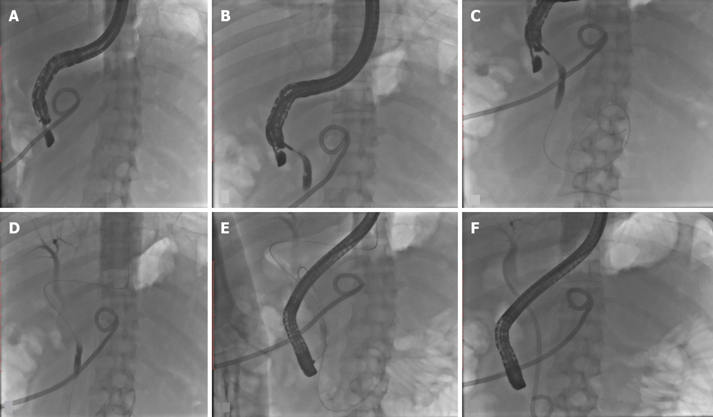
Figure 4 Conventional endoscopic ultrasound guided rendezvous technique (extrahepatic route) being performed in a case of acute biliary pancreatitis (percutaneous catheter drain in situ) diagnosed with choledocholithiasis.
A: Endoscopic ultrasound (EUS) scope positioned in short scope position in D1-D2 junction; B: After common bile duct puncture (access diameter 43 mm) using 19 G EUS- fine needle aspiration needle, bile aspiration was done to confirm position and thereafter, contrast was injected to delineate the bile duct; C: Guidewire was passed across the papilla and coiled in the duodenum; D: EUS scope was exchanged with guidewire in place; E: Subsequent endoscopic retrograde cholangiopancreatography was performed using a duodenoscope and successful biliary cannulation was achieved along the side of the exiting guidewire; F: After stone extraction using balloon sweeps, common bile duct stent (7 French × 7 cm double pigtail stent) was placed.
- Citation: Mishra S, Dhar J, Crinò SF, Samanta J. Technical overview of difficult biliary cannulation in endoscopic retrograde cholangiopancreatography: A guide for the endoscopist. World J Gastroenterol 2026; 32(26): 114936
- URL: https://www.wjgnet.com/1007-9327/full/v32/i26/114936.htm
- DOI: https://dx.doi.org/10.3748/wjg.114936