
















Copyright: ©Author(s) 2026.
World J Gastroenterol. Jul 14, 2026; 32(26): 114936
Published online Jul 14, 2026. doi: 10.3748/wjg.114936
Published online Jul 14, 2026. doi: 10.3748/wjg.114936
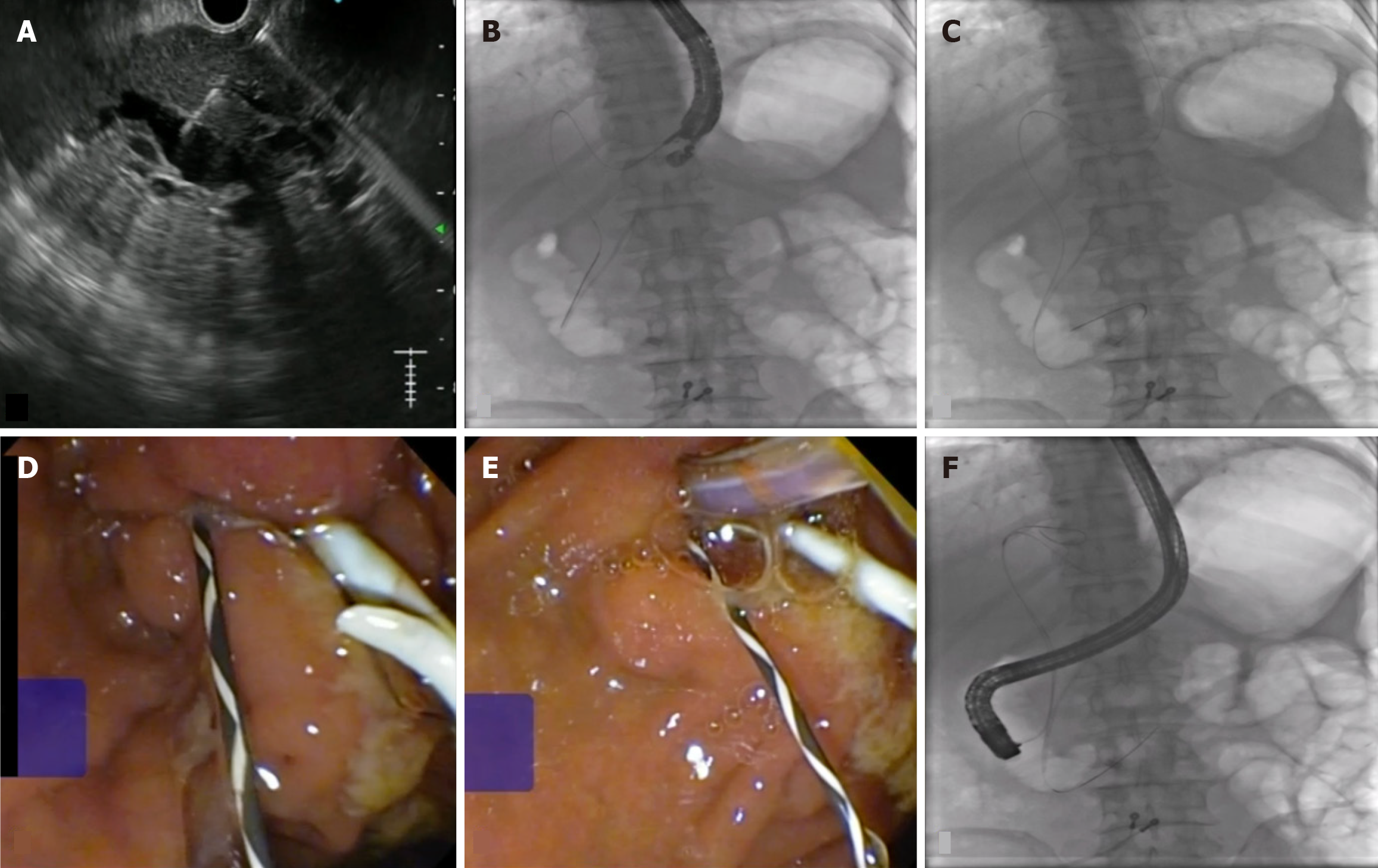
Figure 3 Conventional endoscopic ultrasound guided rendezvous technique (intrahepatic route): A: Endoscopic ultrasound (EUS) image showing dilated intrahepatic biliary radicle and puncture made using 19-G EUS-fine needle aspiration needle; B: After bile aspiration and confirming bile duct puncture, guidewire passed across the papilla into the duodenum as shown in the fluoroscopic image; C: After coiling the guidewire in the duodenum, the EUS scope was exchanged over the guidewire; D: Endoscopic image of the papilla showing a straight plastic stent placed in pancreatic duct (at the time of difficult biliary cannulation during conventional endoscopic retrograde cholangiopancreatography) and the guidewire in the bile duct passed during EUS-rendezvous procedure; E: Selective cannulation of bile duct performed using a sphincterotome by the side of the guidewire; F: Fluoroscopic image showing deep cannulation of the bile duct during endoscopic retrograde cholangiopancreatography procedure; also noted is the guidewire within the bile duct (placed during EUS-rendezvous procedure) with pancreatic duct stent in place.
- Citation: Mishra S, Dhar J, Crinò SF, Samanta J. Technical overview of difficult biliary cannulation in endoscopic retrograde cholangiopancreatography: A guide for the endoscopist. World J Gastroenterol 2026; 32(26): 114936
- URL: https://www.wjgnet.com/1007-9327/full/v32/i26/114936.htm
- DOI: https://dx.doi.org/10.3748/wjg.114936