
















Copyright: ©Author(s) 2026.
World J Gastroenterol. Jul 7, 2026; 32(25): 116783
Published online Jul 7, 2026. doi: 10.3748/wjg.116783
Published online Jul 7, 2026. doi: 10.3748/wjg.116783
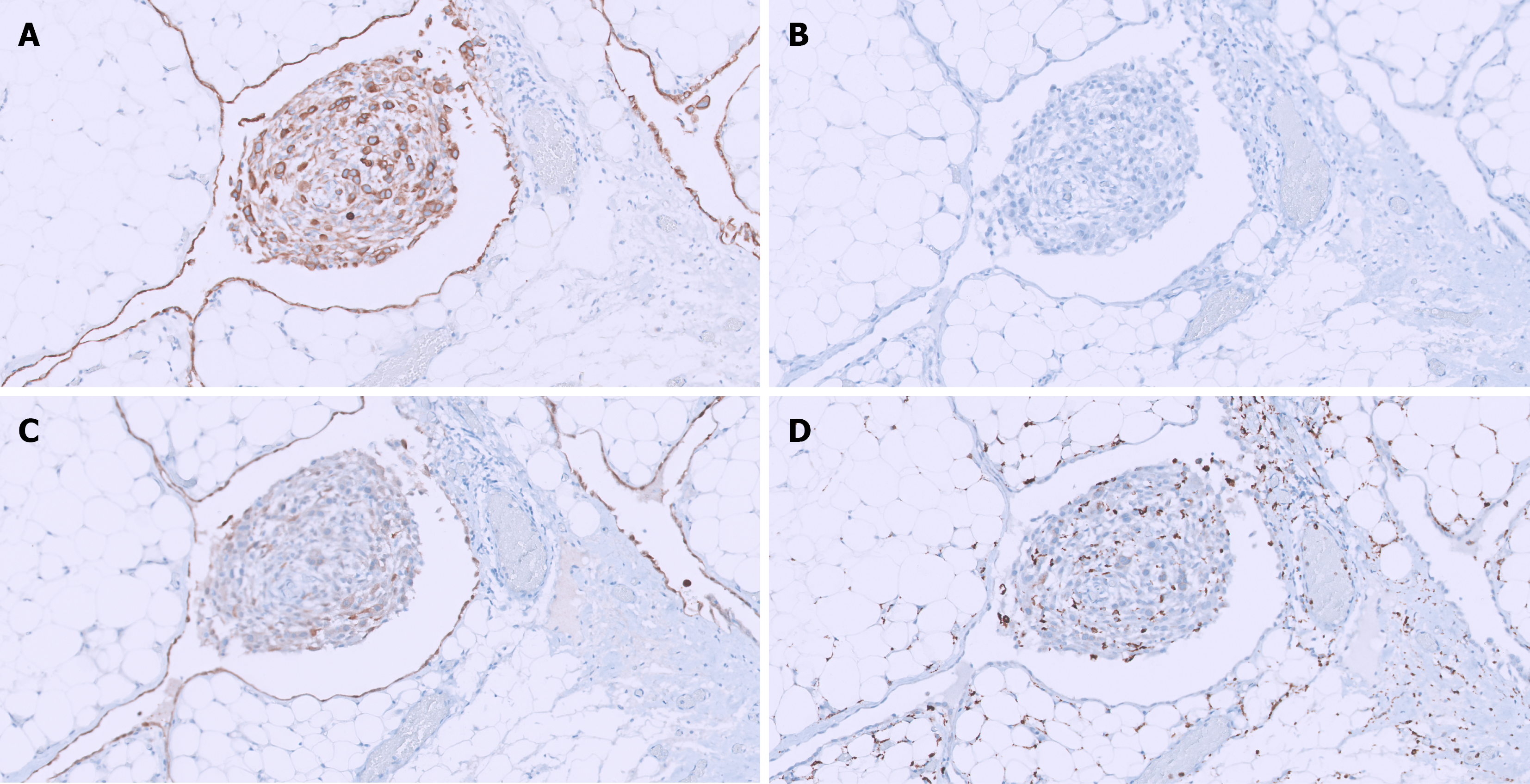
Figure 3 Immunohistochemical findings.
A: Immunohistochemical analyses showing cytokeratin AE1/AE3 positivity in both epithelial and mesothelial cell components [immunohistochemistry (IHC); original magnification 200 ×]; B: Intestinal differentiation marker CDX2 mildly expressed in sparse nuclei of neoplastic cells, indicating a gastric origin (IHC; original magnification 200 ×); C: Immunoexpression of calretinin in mesothelial cells (IHC; original magnification 200 ×); D: Note CD68 immunopositivity highlighting the histiocytic component of nodular histiocytic/mesothelial hyperplasia, while mesothelial cells are negative (IHC; original magnification 200 ×).
- Citation: Colombo P, Dani G, Saracini B, Castoro C, Spaggiari P. Abdominal nodular histiocytic/mesothelial hyperplasia - intraoperative pitfalls, differential diagnosis, literature review: A case report. World J Gastroenterol 2026; 32(25): 116783
- URL: https://www.wjgnet.com/1007-9327/full/v32/i25/116783.htm
- DOI: https://dx.doi.org/10.3748/wjg.116783