
















Copyright: ©Author(s) 2026.
World J Gastroenterol. Jul 7, 2026; 32(25): 116783
Published online Jul 7, 2026. doi: 10.3748/wjg.116783
Published online Jul 7, 2026. doi: 10.3748/wjg.116783
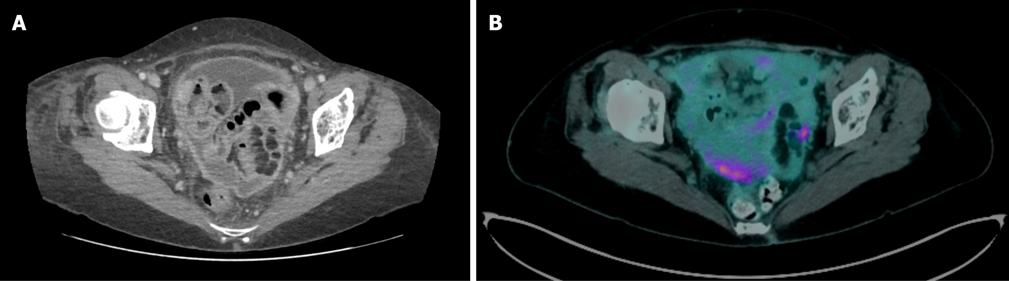
Figure 1 Imaging findings.
A: Computed tomography scan documented the presence of a multifocal thickening of the peritoneum, with confluent nodules visible in the pelvic cavity; B: Positron emission tomography-computed tomography scan showing uptake of [18F]-fluorodeoxyglucose in the left pericolic region and pelvic cavity, suspicious for peritoneal carcinomatosis.
- Citation: Colombo P, Dani G, Saracini B, Castoro C, Spaggiari P. Abdominal nodular histiocytic/mesothelial hyperplasia - intraoperative pitfalls, differential diagnosis, literature review: A case report. World J Gastroenterol 2026; 32(25): 116783
- URL: https://www.wjgnet.com/1007-9327/full/v32/i25/116783.htm
- DOI: https://dx.doi.org/10.3748/wjg.116783