
















Copyright: ©Author(s) 2026.
World J Gastroenterol. Jun 28, 2026; 32(24): 119006
Published online Jun 28, 2026. doi: 10.3748/wjg.119006
Published online Jun 28, 2026. doi: 10.3748/wjg.119006
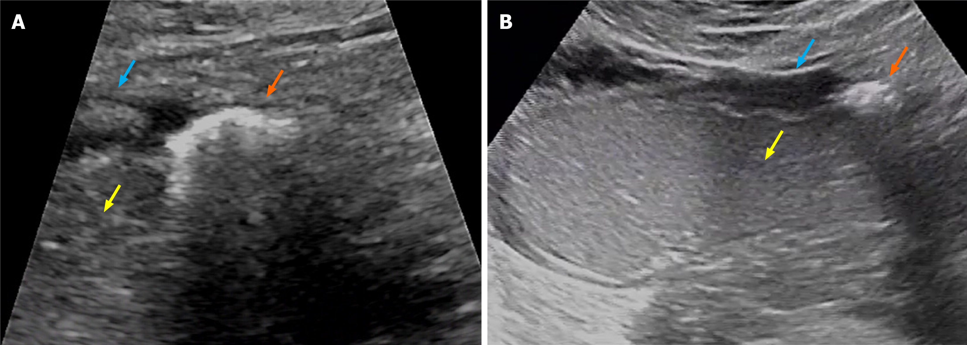
Figure 3 Oral agent contrast-enhanced ultrasound findings.
A: The short-axis section; B: The long-axis section. The thickened and edematous gastric wall surrounding the chronic gastric ulcer is clearly visible, with distinct layers discernible. The ulceration and its attachments extend from the mucosal layer to the serosal layer. The typical edematous hypoechoic base of the ulcer is absent, appearing only as a blurred linear hypoechoic line. Orange arrow: Ulcer; Blue arrow: Thickened gastric wall; Yellow arrow: Gastric lumen.
- Citation: Wei ML, Tan QL. Chronic ulcer diagnosis and perforation risk with oral agent contrast-enhanced ultrasound: A case report and review of literature. World J Gastroenterol 2026; 32(24): 119006
- URL: https://www.wjgnet.com/1007-9327/full/v32/i24/119006.htm
- DOI: https://dx.doi.org/10.3748/wjg.119006