
















Copyright: ©Author(s) 2026.
World J Gastroenterol. Jun 21, 2026; 32(23): 118826
Published online Jun 21, 2026. doi: 10.3748/wjg.v32.i23.118826
Published online Jun 21, 2026. doi: 10.3748/wjg.v32.i23.118826
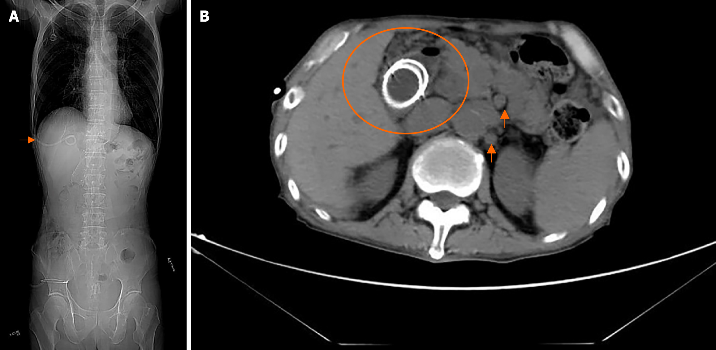
Figure 2 Admission computed tomography.
A: Percutaneous transhepatic biliary drainage catheter (arrow); B: Deformation and luminal narrowing of the descending duodenal stent (circle) and the hepatogastric hiatus and multiple small retroperitoneal lymph nodes (arrow).
- Citation: Guo CY, Wei YX, Li J, Ren GH, Cao J, Sun L, Zhao JY. Percutaneous transhepatic biliary drainage guided endoscopic retrograde cholangiopancreatography rendezvous technique for biliary stent placement: A case report. World J Gastroenterol 2026; 32(23): 118826
- URL: https://www.wjgnet.com/1007-9327/full/v32/i23/118826.htm
- DOI: https://dx.doi.org/10.3748/wjg.v32.i23.118826