
















Copyright: ©Author(s) 2026.
World J Gastroenterol. Jun 14, 2026; 32(22): 118810
Published online Jun 14, 2026. doi: 10.3748/wjg.v32.i22.118810
Published online Jun 14, 2026. doi: 10.3748/wjg.v32.i22.118810
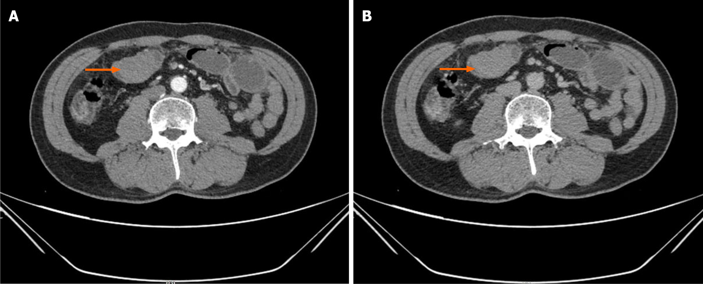
Figure 1 Contrast-enhanced abdominal computed tomography scan showing small intestinal mass.
A: Arterial phase axial image showing a well-defined enhancing mass (arrow) in the right mid-abdomen, causing proximal small bowel dilation; B: Portal venous phase confirms the soft-tissue density mass (arrow) with associated small bowel obstruction, initially suspected to be a gastrointestinal stromal tumor.
- Citation: Lin JQ, Liu HZ, Li ZW, Yang K. Intestinal myeloid sarcoma mimicking gastrointestinal stromal tumor without leukemic manifestations: A case report and review of literature. World J Gastroenterol 2026; 32(22): 118810
- URL: https://www.wjgnet.com/1007-9327/full/v32/i22/118810.htm
- DOI: https://dx.doi.org/10.3748/wjg.v32.i22.118810