
















Copyright: ©Author(s) 2026.
World J Gastroenterol. Jun 14, 2026; 32(22): 117194
Published online Jun 14, 2026. doi: 10.3748/wjg.v32.i22.117194
Published online Jun 14, 2026. doi: 10.3748/wjg.v32.i22.117194
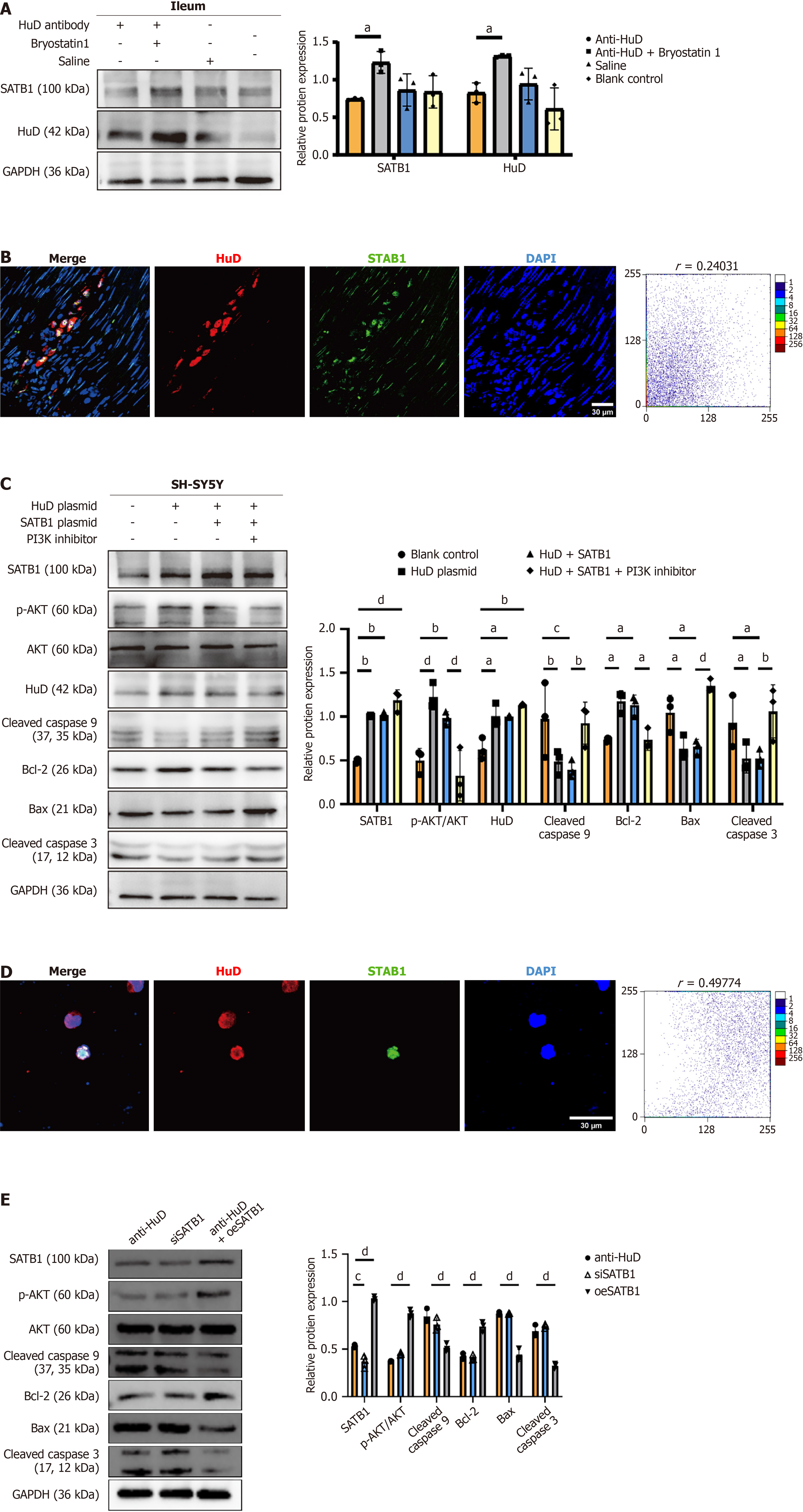
Figure 7 Anti-HuD autoantibody induced neuronal apoptosis through the special AT-rich sequence-binding protein 1-phosphatidy
- Citation: Zhang Y, Zhu C, Xia SH, Zhang MY, Liu YR, Guan JL, Huang YJ, Zhao K, Liao JZ, Fan WJ. Mechanism of anti-human antigen D (HuD/ELAVL4) autoantibody-induced enteric neuronal apoptosis in irritable bowel syndrome and its potential for targeted intervention. World J Gastroenterol 2026; 32(22): 117194
- URL: https://www.wjgnet.com/1007-9327/full/v32/i22/117194.htm
- DOI: https://dx.doi.org/10.3748/wjg.v32.i22.117194