
















Copyright: ©Author(s) 2026.
World J Gastroenterol. Jun 14, 2026; 32(22): 117194
Published online Jun 14, 2026. doi: 10.3748/wjg.v32.i22.117194
Published online Jun 14, 2026. doi: 10.3748/wjg.v32.i22.117194
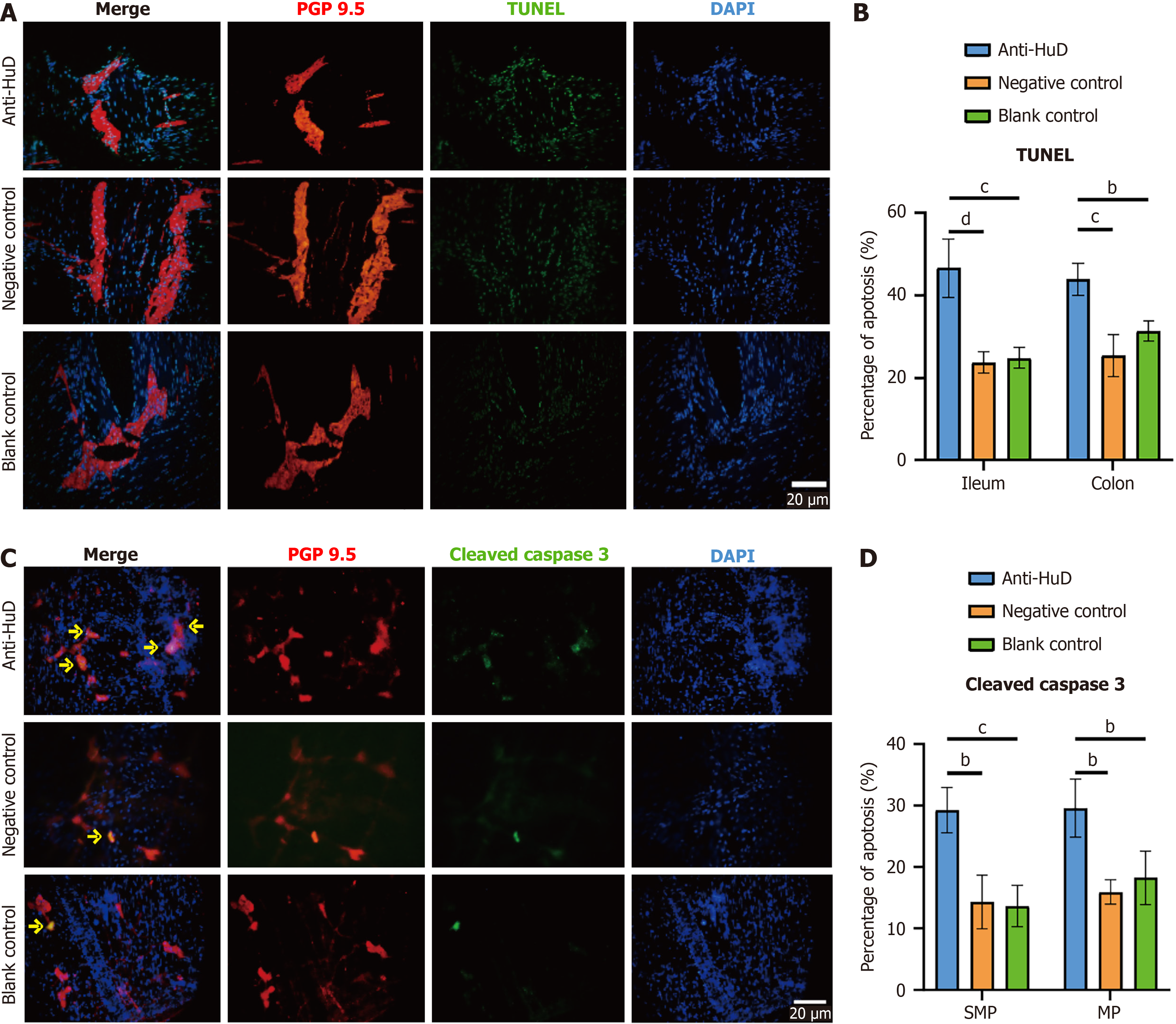
Figure 4 Anti-HuD autoantibody induced neuronal apoptosis in an irritable bowel syndrome rat model.
A: Terminal-deoxynucleotidyl transferase mediated nick end labeling of neurons in the ileal plexus of the three groups of rats with neurons indicated by PGP 9.5; B: Cleaved caspase 3 staining of neurons in the ileal plexus of the three groups of rats with enteric neurons indicated by PGP 9.5; C: Percentage of apoptotic neurons by terminal-deoxynucleotidyl transferase mediated nick end labeling; D: Percentage of apoptotic neurons by cleaved caspase 3 staining. The yellow arrows indicate apoptotic enteric neurons. Scale bar: 20 μm. Data are expressed as mean ± SD. C and D were analyzed by two-way analysis of variance with Tukey’s multiple comparison test, and n = 3 for each group. bP < 0.01, cP < 0.001, and dP < 0.0001. DAPI: 4’,6-diamidino-2-phenylindole; TUNEL: Terminal-deoxynucleotidyl transferase mediated nick end labeling.
- Citation: Zhang Y, Zhu C, Xia SH, Zhang MY, Liu YR, Guan JL, Huang YJ, Zhao K, Liao JZ, Fan WJ. Mechanism of anti-human antigen D (HuD/ELAVL4) autoantibody-induced enteric neuronal apoptosis in irritable bowel syndrome and its potential for targeted intervention. World J Gastroenterol 2026; 32(22): 117194
- URL: https://www.wjgnet.com/1007-9327/full/v32/i22/117194.htm
- DOI: https://dx.doi.org/10.3748/wjg.v32.i22.117194