
















Copyright: ©Author(s) 2026.
World J Gastroenterol. Jun 14, 2026; 32(22): 117194
Published online Jun 14, 2026. doi: 10.3748/wjg.v32.i22.117194
Published online Jun 14, 2026. doi: 10.3748/wjg.v32.i22.117194
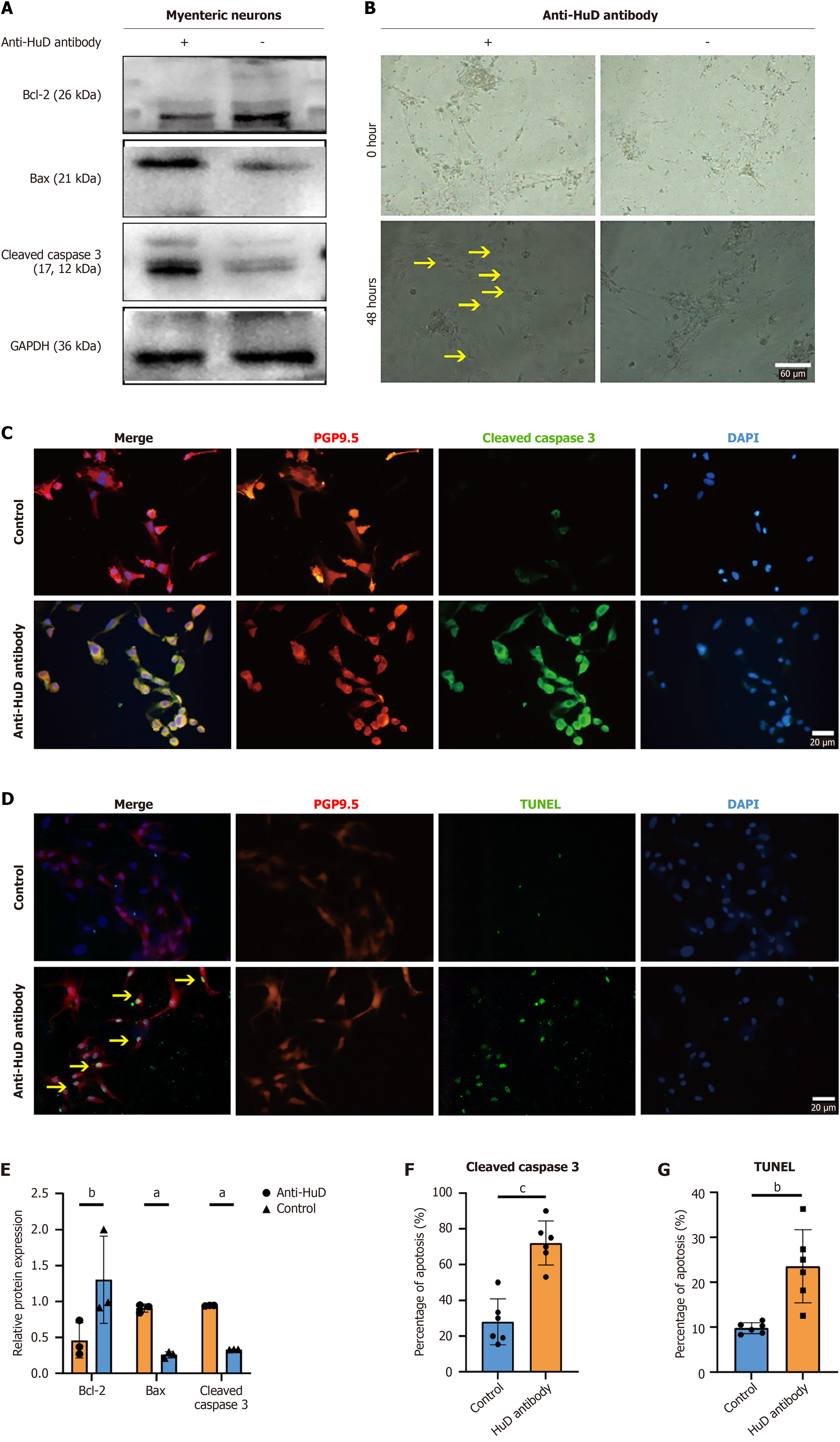
Figure 2 Anti-HuD autoantibody induced apoptosis of primary enteric neurons.
A: Expression of apoptotic proteins detected by western blot; B: Morphological changes after 48 hours of antibody incubation. More apoptotic cells with vacant cytoplasm, partial atrophy, and reduced refraction were observed in the treated group. The yellow arrows indicate apoptotic neurons. Scale bar: 60 μm; C: Immunofluorescence detected the expression of cleaved caspase 3, and anti-PGP 9.5 indicated neurons; D: Immunofluorescence and terminal-deoxynucleotidyl transferase mediated nick end labeling staining detected nuclei cleavage. Anti-PGP 9.5 indicated neurons. The yellow arrows indicate apoptotic neurons. Scale bar: 20 μm; E: Statistical analysis of the relative protein expression of Bcl-2, Bax, and cleaved caspase 3; F: Statistical analysis of the percentage of apoptosis using cleaved caspase 3 immunostaining; G: Statistical analysis of the percentage of apoptosis using terminal-deoxynucleotidyl transferase mediated nick end labeling immunostaining. Data are expressed as mean ± SD; Student’s t-test was performed, n = 6 for each group. aP < 0.05, bP < 0.01, cP < 0.001. DAPI: 4’,6-diamidino-2-phenylindole; GAPDH: Glyceraldehyde-3-phosphate dehydrogenase; TUNEL: Terminal-deoxynucleotidyl transferase mediated nick end labeling.
- Citation: Zhang Y, Zhu C, Xia SH, Zhang MY, Liu YR, Guan JL, Huang YJ, Zhao K, Liao JZ, Fan WJ. Mechanism of anti-human antigen D (HuD/ELAVL4) autoantibody-induced enteric neuronal apoptosis in irritable bowel syndrome and its potential for targeted intervention. World J Gastroenterol 2026; 32(22): 117194
- URL: https://www.wjgnet.com/1007-9327/full/v32/i22/117194.htm
- DOI: https://dx.doi.org/10.3748/wjg.v32.i22.117194