
















Copyright: ©Author(s) 2026.
World J Gastroenterol. May 28, 2026; 32(20): 116020
Published online May 28, 2026. doi: 10.3748/wjg.v32.i20.116020
Published online May 28, 2026. doi: 10.3748/wjg.v32.i20.116020
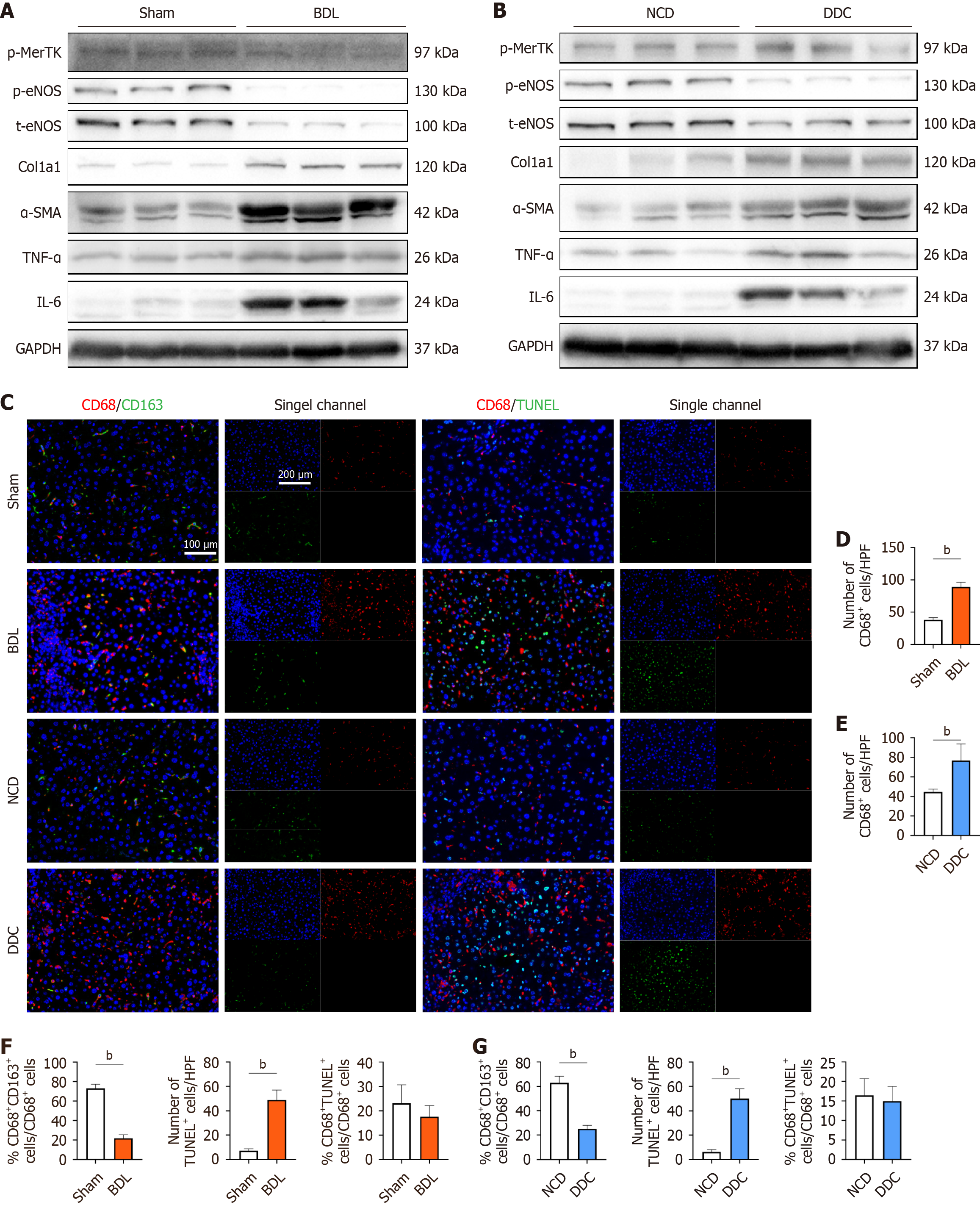
Figure 2 Efferocytosis was defective in mice models of cholestasis and portal hypertension.
A: Western blotting analysis of MerTK phosphorylation, intrahepatic vascular resistance (IHVR) (p-eNOS and t-eNOS), fibrosis (Col1a1 and α-SMA), and inflammation [tumor necrosis factor α (TNF-α) and interleukin (IL)-6] in liver from sham and bile duct ligation (BDL) mice; B: Western blotting analysis of MerTK phosphorylation, IHVR (p-eNOS and t-eNOS), fibrosis (Col1a1 and α-SMA), and inflammation (TNF-α and IL-6) in liver from normal chow diet (NCD) and 3,5-diethoxycarbonyl-1,4-dihydrocollidine (DDC) mice; C: The representative images of liver sections probed with specific antibodies against the macrophage marker CD68 and reparative macrophage marker CD163, or with a co-staining of TUNEL assay; D: The quantification of macrophage infiltration in sham and BDL mice; E: The quantification of macrophage infiltration in NCD and DDC mice; F: The quantifications of reparative macrophages, apoptotic cells, and macrophage efferocytosis in sham and BDL mice; G: The quantifications of reparative macrophages, apoptotic cells, and efferocytosis in NCD and DDC mice. bP < 0.01. NCD: Normal chow diet; DDC: Diethoxycarbonyl-1,4-dihydrocollidine; BDL: Bile duct ligation; TNF-α: Tumor necrosis factor α; IL: Interleukin.
- Citation: Luo GQ, Zhao JB, Wu ZH, Lin JY, Zhang CH, Wu GB, Fan Q, Qi XL, Li HJ, Luo M, Zheng L. Growth arrest specific 6 ameliorated inflammation and portal pressure through activating efferocytosis in cholestasis and portal hypertension. World J Gastroenterol 2026; 32(20): 116020
- URL: https://www.wjgnet.com/1007-9327/full/v32/i20/116020.htm
- DOI: https://dx.doi.org/10.3748/wjg.v32.i20.116020