
















©The Author(s) 2026.
World J Gastroenterol. Jan 14, 2026; 32(2): 114222
Published online Jan 14, 2026. doi: 10.3748/wjg.v32.i2.114222
Published online Jan 14, 2026. doi: 10.3748/wjg.v32.i2.114222
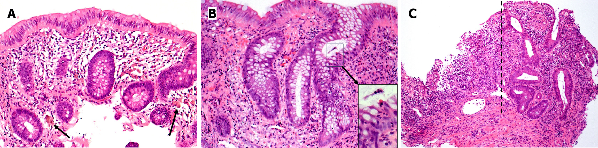
Figure 2 Microscopic photographs of endoscopic biopsies (obtained at age 40 while managing ulcerative colitis with only oral corticosteroids), hematoxylin and eosin stained histologic sections.
A: Right colon biopsy (medium power microscopic view photographed at 100 × magnification) reveals colonic mucosa with occasional plump macrophages with pigmented cytoplasmic accumulation (arrows) within the lamina propria. Colonic mucosa is, otherwise, histologically unremarkable; B: Left colon biopsy (medium power microscopic view photographed at 100 × magnification) reveals mildly active chronic colitis with crypt architectural distortion and a mild increase in mononuclear inflammatory cells within the lamina propria. The epithelium in the crypt on the far right is infiltrated by several neutrophils (“cryptitis”) with modest infiltration into the crypt of Lieberkühn (insert: High power microscopic view); C: Rectal biopsy (low power microscopic view photographed at 40 × magnification) reveals active chronic colitis with crypt architectural distortion and ulceration. The mucosa on the right (of the dotted line) contains a moderate increase in mononuclear inflammatory cells within the lamina propria, basal plasmacytosis, and crypt architectural distortion; whereas, the mucosa on the left (of the dotted line) is ulcerated, comprised of inflamed granulation tissue without crypts.
- Citation: Sylvestre PB. Reducing agents for induction and maintenance therapy achieve long-term remission of refractory ulcerative colitis: A case report and review of literature. World J Gastroenterol 2026; 32(2): 114222
- URL: https://www.wjgnet.com/1007-9327/full/v32/i2/114222.htm
- DOI: https://dx.doi.org/10.3748/wjg.v32.i2.114222