
















©The Author(s) 2026.
World J Gastroenterol. Jan 14, 2026; 32(2): 114222
Published online Jan 14, 2026. doi: 10.3748/wjg.v32.i2.114222
Published online Jan 14, 2026. doi: 10.3748/wjg.v32.i2.114222
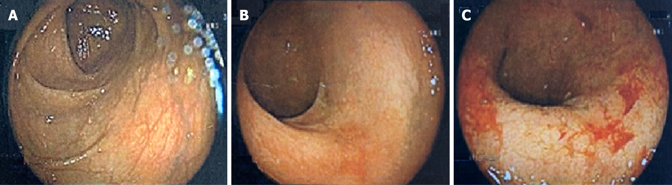
Figure 1 Colonoscopy images (colonoscopy performed at age 40 while managing ulcerative colitis with only oral corticosteroids).
A: Proximal left colon with normal vascular pattern and no mucosal lesions; B: Sigmoid colon with erythema and mucosal friability; C: Rectum with erythema, mucosal friability, erosions, and ulceration.
- Citation: Sylvestre PB. Reducing agents for induction and maintenance therapy achieve long-term remission of refractory ulcerative colitis: A case report and review of literature. World J Gastroenterol 2026; 32(2): 114222
- URL: https://www.wjgnet.com/1007-9327/full/v32/i2/114222.htm
- DOI: https://dx.doi.org/10.3748/wjg.v32.i2.114222