
















Copyright: ©Author(s) 2026.
World J Gastroenterol. May 21, 2026; 32(19): 116837
Published online May 21, 2026. doi: 10.3748/wjg.v32.i19.116837
Published online May 21, 2026. doi: 10.3748/wjg.v32.i19.116837
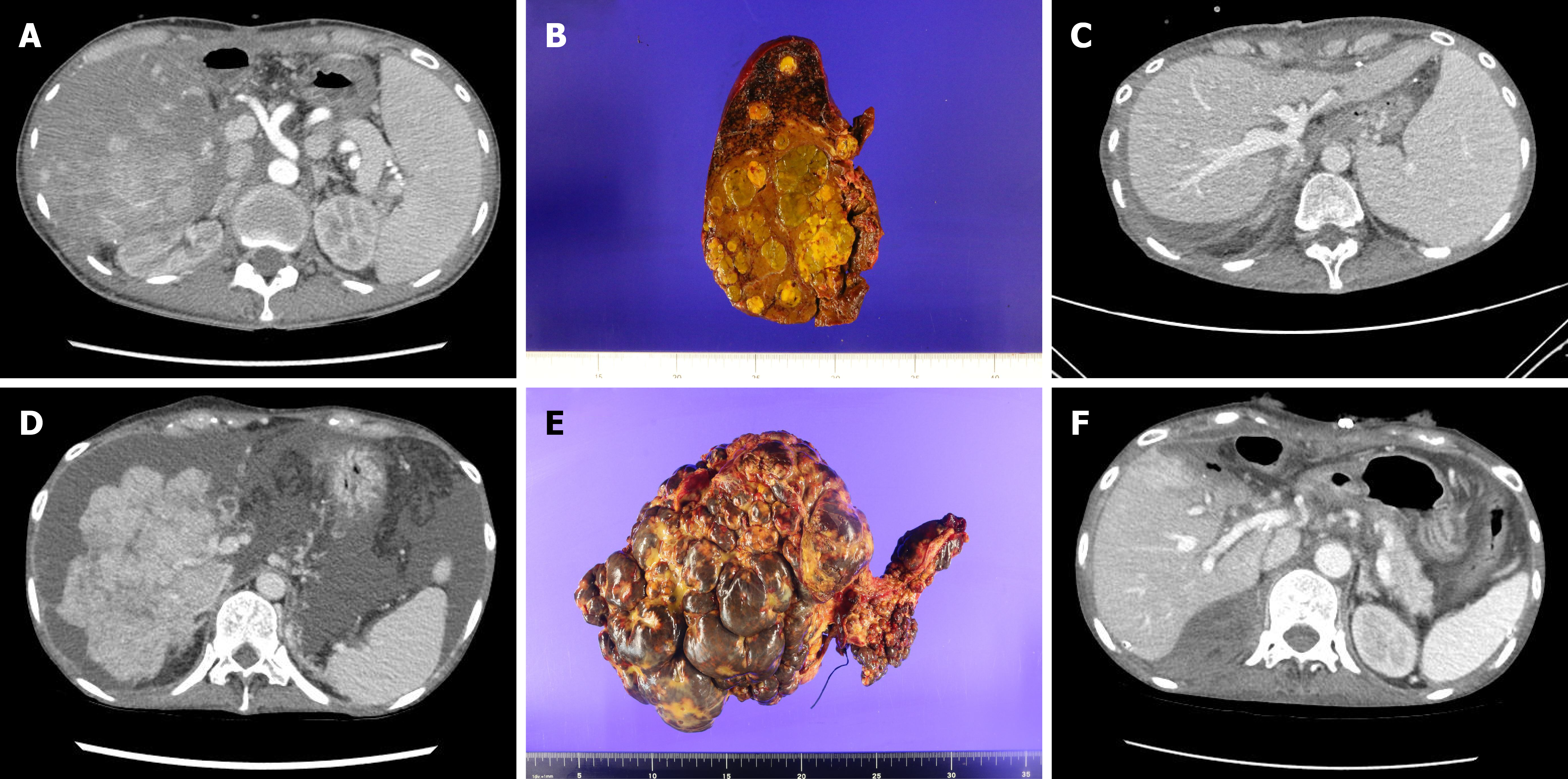
Figure 1 Pre- and post-transplant imaging and explant specimens in two patients undergoing rescue liver transplantation.
A: Pre-transplant contrast-enhanced computed tomography (CECT) of case 1 showing multifocal hepatic tumor burden; B: Explanted liver from case 1 demonstrating multiple well-circumscribed yellowish nodules consistent with treated and viable hepatoblastoma; C: Post-transplant CECT of case 1 showing normal graft enhancement; D: Pre-transplant CECT of case 2 showing diffuse metastatic involvement of the liver; E: Explanted liver from case 2 demonstrating diffuse bosselated metastatic replacement; F: Post-transplant CECT of case 2 showing homogeneous graft enhancement.
- Citation: Kim SH, An BH, Lee JA, Jeong GW. Rescue liver transplantation for liver failure with extrahepatic metastases: An ethical and clinical framework informed by two cases. World J Gastroenterol 2026; 32(19): 116837
- URL: https://www.wjgnet.com/1007-9327/full/v32/i19/116837.htm
- DOI: https://dx.doi.org/10.3748/wjg.v32.i19.116837