
















Copyright: ©Author(s) 2026.
World J Gastroenterol. May 21, 2026; 32(19): 113022
Published online May 21, 2026. doi: 10.3748/wjg.v32.i19.113022
Published online May 21, 2026. doi: 10.3748/wjg.v32.i19.113022
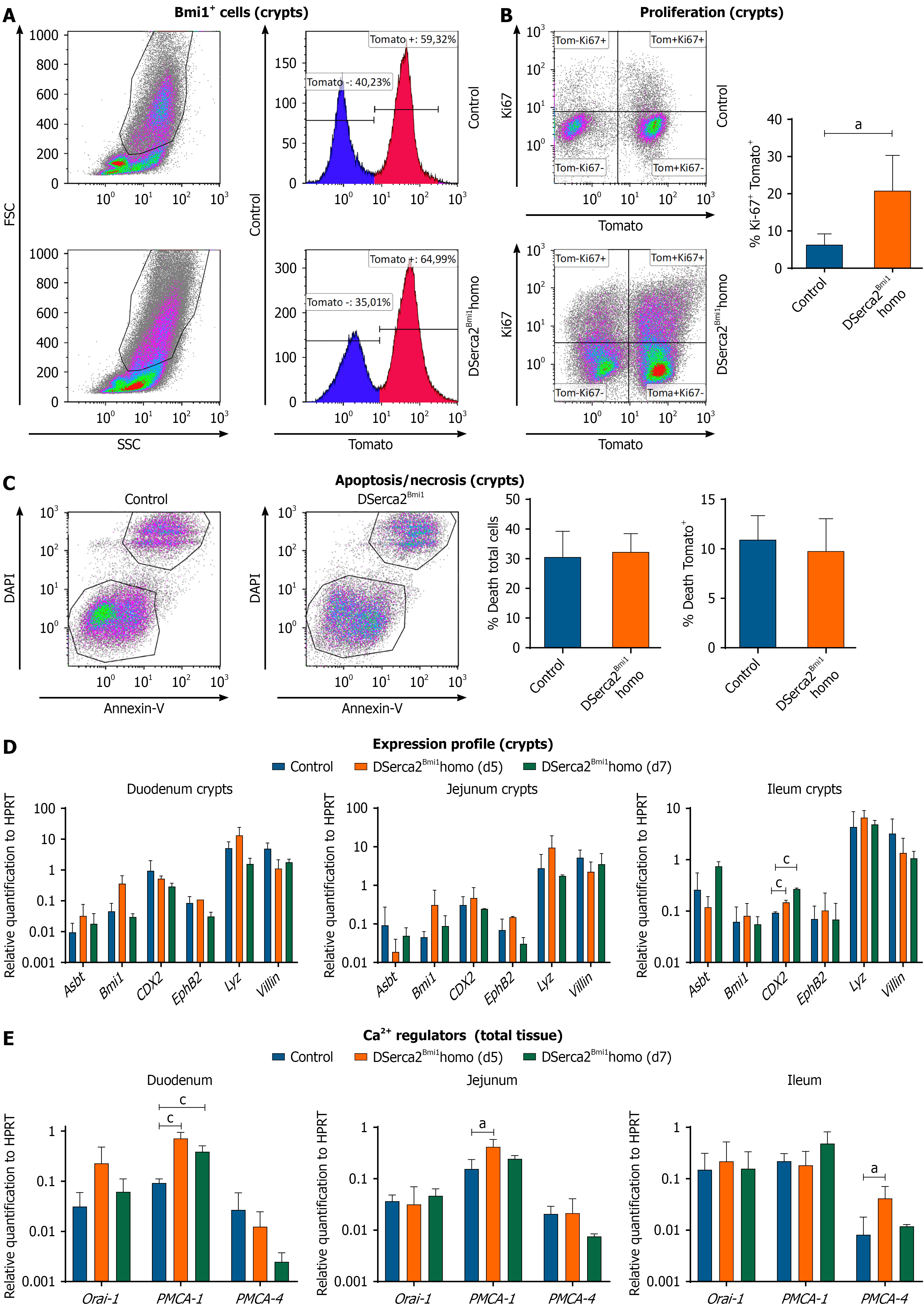
Figure 3 Impact of conditional Serca2 deletion in the small intestine.
A-C: Analysis of crypt-enriched fractions isolated in parallel from DSerca2Bmi1homo and control animals [5 days post-tamoxifen (Tx)]. A: Estimation of Bmi1+ cells (Tmt+) by flow cytometry (n = 3 each; independent animals); B: Proliferation analysis (Ki-67+) by flow cytometry (n = 3 each; independent animals); C: Evaluation of apoptosis/necrosis rate by flow cytometry (4’,6-diamidino-2-phenylindole/Annexin-V) (n = 3 each; independent animals); D and E: Expression analysis (real-time quantitative polymerase chain reaction) in small intestine segments (duodenum; jejunum; ileum) from DSerca2Bmi1homo and control mice; Control (n = 5 each; independent animals) and DSerca2Bmi1homo 5 days (n = 3 each; independent animals) and 7 days (n = 2 each; independent animals) mice. All samples were normalized to levels of the housekeeping gene Hprt. Analysis of a panel of functionally relevant genes in intestinal crypts in crypt-enriched fractions of the indicated animals (n = 3 each; independent animals). D: Analysis of a panel of relevant genes involved in calcium ions regulation in total tissue of the indicated groups of animals (n = 3 each; independent animals); E: Assays were performed three times and data are expressed as mean ± SD. aP < 0.05. cP < 0.001. FSC: Forward scatter; SSC: Side scatter; DAPI: 4’,6-diamidino-2-phenylindole; Ca2+: Calcium ions.
- Citation: Herrero D, Docavo A, Salvador M, Higuera M, Cortés A, García-Ceca J, Montero-Herradón S, García-Brenes MA, Albericio G, Cordero A, Carmona RM, de Molina A, Salvador JM, Mora C, Zapata AG, Bernad A. Serca2 deletion in the mouse adult Bmi1+ compartment induces a lethal phenotype involving a severe gastric dysfunction. World J Gastroenterol 2026; 32(19): 113022
- URL: https://www.wjgnet.com/1007-9327/full/v32/i19/113022.htm
- DOI: https://dx.doi.org/10.3748/wjg.v32.i19.113022