
















Copyright: ©Author(s) 2026.
World J Gastroenterol. May 21, 2026; 32(19): 112955
Published online May 21, 2026. doi: 10.3748/wjg.v32.i19.112955
Published online May 21, 2026. doi: 10.3748/wjg.v32.i19.112955
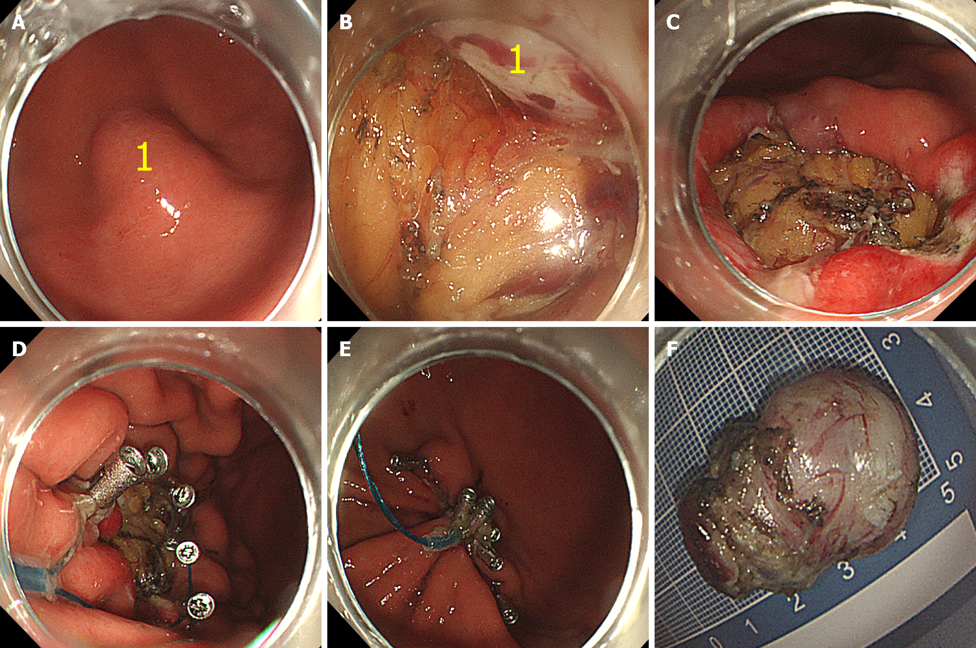
Figure 1 Conventional endoscopic full-thickness resection technology.
A: Subepithelial tumor located on the greater curvature of the lower stomach; B and C: Tumor exposure achieved with a transparent cap to delineate the interface between the lesion and surrounding tissue, followed by resection, leaving a large gastric wall defect; D and E: Defect closed using purse-string suture with clips and an endoloop ligation device; F: Resected specimen showing en bloc removal. 1: Indicates the tumor.
- Citation: Li X, Zhang RY, Wen XD, Li XQ, Liu WH. Traction-preclosure-assisted vs conventional endoscopic full-thickness resection for gastric subepithelial tumors: Safety, efficacy in a retrospective cohort (with video). World J Gastroenterol 2026; 32(19): 112955
- URL: https://www.wjgnet.com/1007-9327/full/v32/i19/112955.htm
- DOI: https://dx.doi.org/10.3748/wjg.v32.i19.112955