
















Copyright: ©Author(s) 2026.
World J Gastroenterol. May 14, 2026; 32(18): 118499
Published online May 14, 2026. doi: 10.3748/wjg.v32.i18.118499
Published online May 14, 2026. doi: 10.3748/wjg.v32.i18.118499
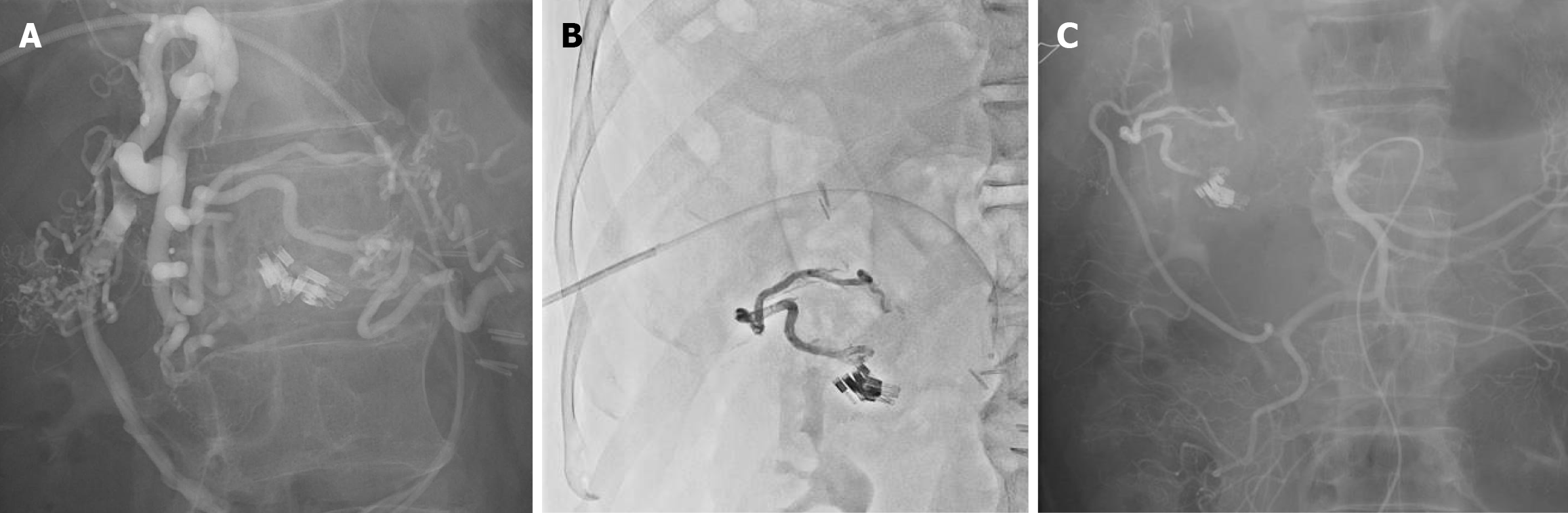
Figure 5 Angiography.
A: The catheter was advanced via the ileocolic vein into the marginal vein, and positioned at the right aspect of the anastomotic varices. Balloon-occluded retrograde venography demonstrated that the variceal drainage extended beyond the previously placed endoscopic clips and drained toward the inferior mesenteric vein; B: Selective occlusion of the two efferent draining veins, achieving unilateral flow control and directing the sclerosing agent into the targeted variceal complex; C: Final angiography demonstrated complete disappearance of the anastomotic varices and elimination of the previously visualized outflow tract, confirming successful obliteration.
- Citation: Tomita D, Yamamoto M, Wakamatsu M, Shibayama R, Maeda Y, Hiramatsu K, Hanaoka Y, Toda S, Ueno M, Matoba S, Kuroyanagi H. Anastomotic colonic varices after colectomy treated by obliteration via a transhepatic portal approach: A case report. World J Gastroenterol 2026; 32(18): 118499
- URL: https://www.wjgnet.com/1007-9327/full/v32/i18/118499.htm
- DOI: https://dx.doi.org/10.3748/wjg.v32.i18.118499