
















Copyright: ©Author(s) 2026.
World J Gastroenterol. May 14, 2026; 32(18): 118499
Published online May 14, 2026. doi: 10.3748/wjg.v32.i18.118499
Published online May 14, 2026. doi: 10.3748/wjg.v32.i18.118499
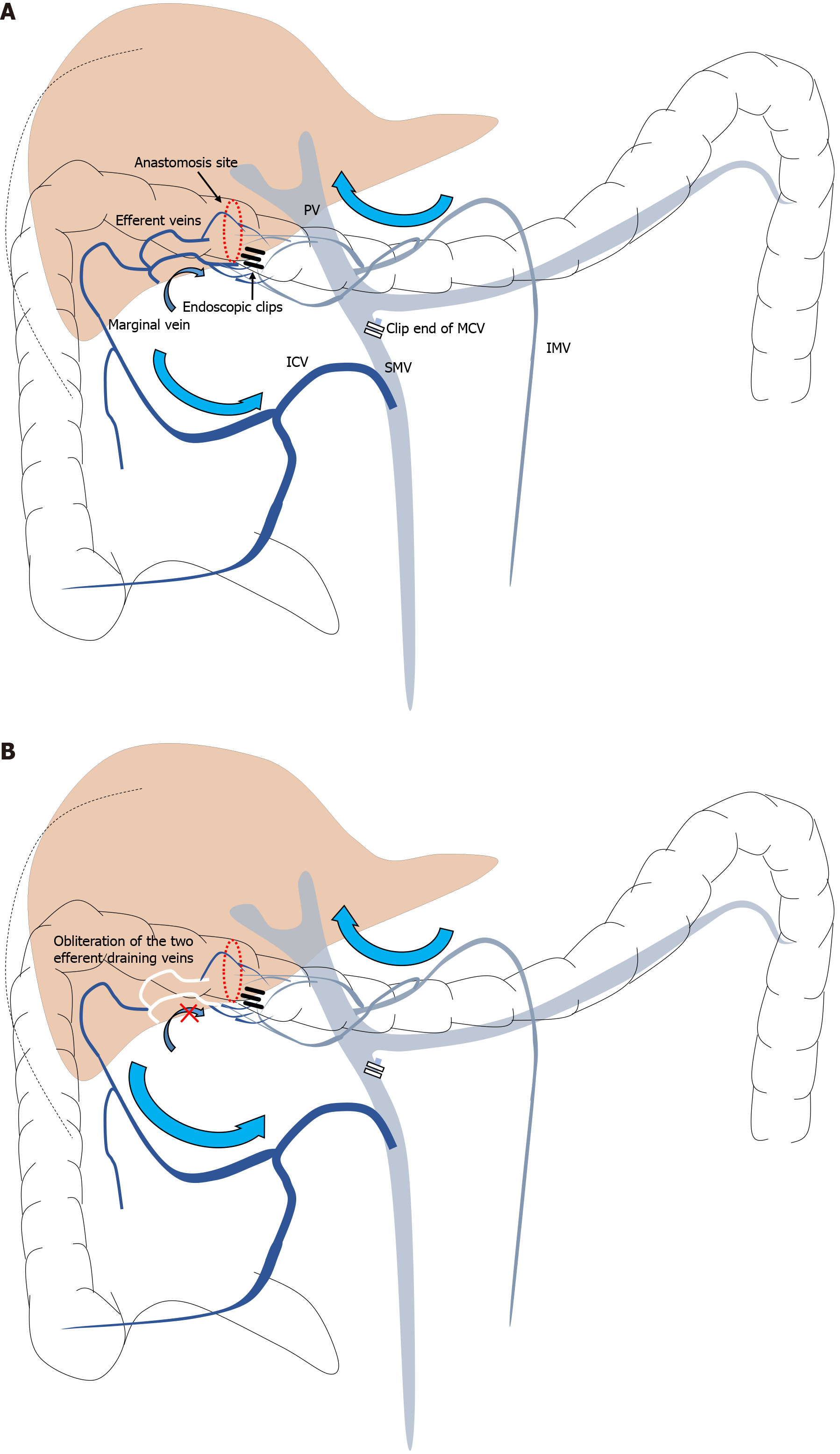
Figure 4 Schematic illustration of venous anatomy.
A: Schematic illustration of venous drainage around the transverse colonic anastomosis after central ligation of the middle colic vein (preembolization). The venous inflow from the marginal vein (superior mesenteric vein territory) converged at the anastomosis site (red dotted circle) and drained into the inferior mesenteric vein via two efferent veins; B: Post-embolization. The two efferent draining veins were selectively embolized with N-butyl-2-cyanoacrylate to establish unilateral flow control, and ethanolamine oleate was injected under balloon occlusion to achieve diffuse thrombosis of the variceal plexus. PV: Portal vein; SMV: Superior mesenteric vein; IMV: Inferior mesenteric vein; ICV: Ileocolic vein; MCV: Middle colic vein.
- Citation: Tomita D, Yamamoto M, Wakamatsu M, Shibayama R, Maeda Y, Hiramatsu K, Hanaoka Y, Toda S, Ueno M, Matoba S, Kuroyanagi H. Anastomotic colonic varices after colectomy treated by obliteration via a transhepatic portal approach: A case report. World J Gastroenterol 2026; 32(18): 118499
- URL: https://www.wjgnet.com/1007-9327/full/v32/i18/118499.htm
- DOI: https://dx.doi.org/10.3748/wjg.v32.i18.118499