
















Copyright: ©Author(s) 2026.
World J Gastroenterol. May 14, 2026; 32(18): 118499
Published online May 14, 2026. doi: 10.3748/wjg.v32.i18.118499
Published online May 14, 2026. doi: 10.3748/wjg.v32.i18.118499
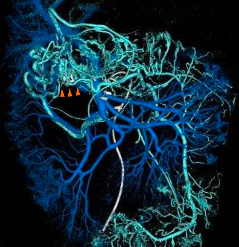
Figure 3 Portal-phase angiography with computed tomography portography.
Portal-phase venography via the superior mesenteric artery (SMA) and inferior mesenteric artery (IMA) demonstrated that transverse colonic varices (orange arrowheads) had developed as collateral venous drainage at the anastomotic site. Blue vessels: Venous return system from the SMA. Light blue vessels: Venous return system from the IMA.
- Citation: Tomita D, Yamamoto M, Wakamatsu M, Shibayama R, Maeda Y, Hiramatsu K, Hanaoka Y, Toda S, Ueno M, Matoba S, Kuroyanagi H. Anastomotic colonic varices after colectomy treated by obliteration via a transhepatic portal approach: A case report. World J Gastroenterol 2026; 32(18): 118499
- URL: https://www.wjgnet.com/1007-9327/full/v32/i18/118499.htm
- DOI: https://dx.doi.org/10.3748/wjg.v32.i18.118499