
















Copyright: ©Author(s) 2026.
World J Gastroenterol. May 14, 2026; 32(18): 118499
Published online May 14, 2026. doi: 10.3748/wjg.v32.i18.118499
Published online May 14, 2026. doi: 10.3748/wjg.v32.i18.118499
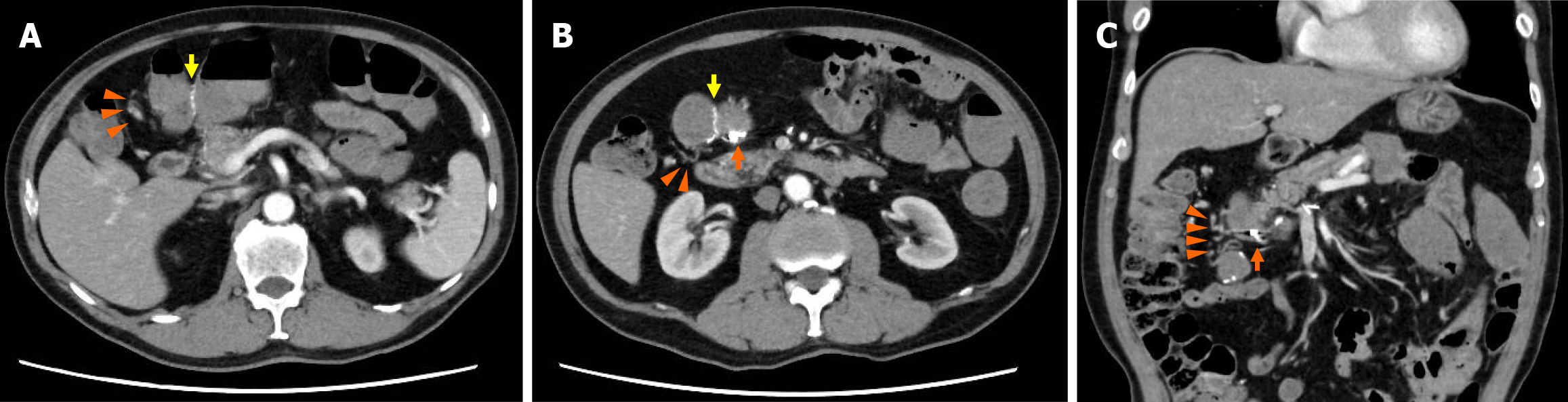
Figure 2 Computed tomography findings.
A and B: Axial contrast-enhanced computed tomography images obtained before the third endoscopic hemostasis procedure. Metallic clips (orange arrow) were observed at the anastomotic site (yellow arrow), with abnormal vascular structures (orange arrowheads) noted around it. A slightly hyperdense intraluminal fluid collection was noted; however, no active extravasation was observed. No imaging findings indicated liver cirrhosis or portal hypertension; C: The coronal view.
- Citation: Tomita D, Yamamoto M, Wakamatsu M, Shibayama R, Maeda Y, Hiramatsu K, Hanaoka Y, Toda S, Ueno M, Matoba S, Kuroyanagi H. Anastomotic colonic varices after colectomy treated by obliteration via a transhepatic portal approach: A case report. World J Gastroenterol 2026; 32(18): 118499
- URL: https://www.wjgnet.com/1007-9327/full/v32/i18/118499.htm
- DOI: https://dx.doi.org/10.3748/wjg.v32.i18.118499