
















Copyright: ©Author(s) 2026.
World J Gastroenterol. May 14, 2026; 32(18): 118499
Published online May 14, 2026. doi: 10.3748/wjg.v32.i18.118499
Published online May 14, 2026. doi: 10.3748/wjg.v32.i18.118499
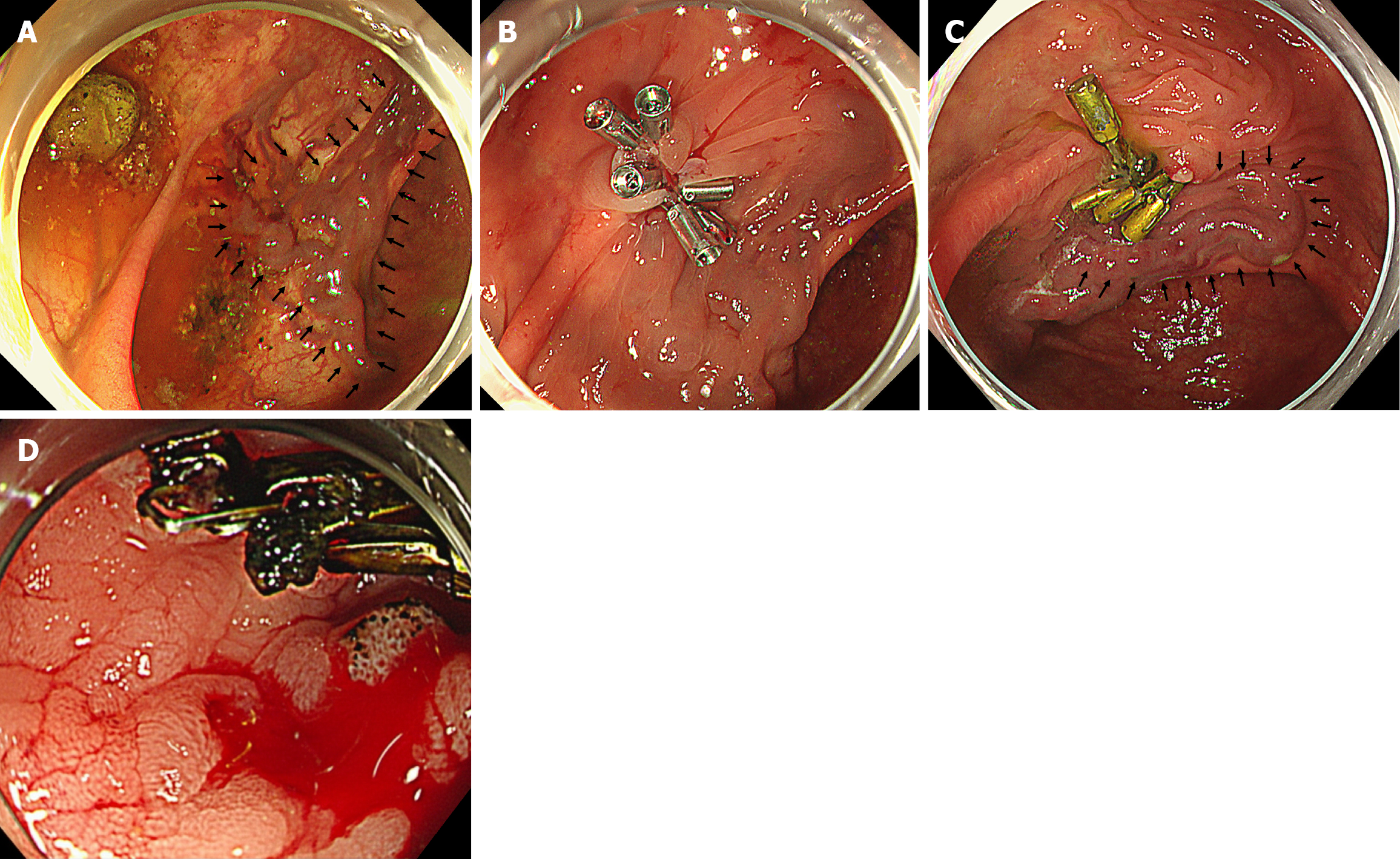
Figure 1 Colonoscopy findings.
A: First endoscopy. Markedly dilated and tortuous submucosal veins (black arrows) around the anastomotic site with active bleeding from the varix; B: Hemostasis was achieved by clipping; C: Second endoscopy. Venous dilatation (black arrows) was observed near the anastomotic site. Exudative bleeding was identified at a location different from the previous clip site; D: Third endoscopy. Recurrent bleeding was observed at a different site from the prior clipping sites.
- Citation: Tomita D, Yamamoto M, Wakamatsu M, Shibayama R, Maeda Y, Hiramatsu K, Hanaoka Y, Toda S, Ueno M, Matoba S, Kuroyanagi H. Anastomotic colonic varices after colectomy treated by obliteration via a transhepatic portal approach: A case report. World J Gastroenterol 2026; 32(18): 118499
- URL: https://www.wjgnet.com/1007-9327/full/v32/i18/118499.htm
- DOI: https://dx.doi.org/10.3748/wjg.v32.i18.118499