
















Copyright: ©Author(s) 2026.
World J Gastroenterol. May 14, 2026; 32(18): 116229
Published online May 14, 2026. doi: 10.3748/wjg.v32.i18.116229
Published online May 14, 2026. doi: 10.3748/wjg.v32.i18.116229
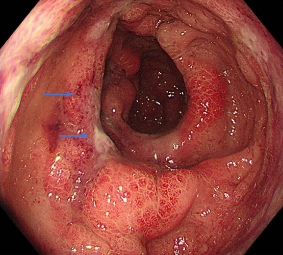
Figure 3 Colonoscopic features of inferior mesenteric vein-related colon ischemia.
Continuous lesions characterized by mucosal congestion and edema, with focal dark purple area, nodular changes and irregular ulcers in distal rectum, as indicated by the arrows.
- Citation: Jin X, Xu TM, Ruan GC, Zhang XF, Chen XY, Li XQ, Tang H, Xu H, Wang QP, Liu W, Li J. Clinical manifestations and imaging features of inferior mesenteric vein-related colon ischemia. World J Gastroenterol 2026; 32(18): 116229
- URL: https://www.wjgnet.com/1007-9327/full/v32/i18/116229.htm
- DOI: https://dx.doi.org/10.3748/wjg.v32.i18.116229