
















Copyright: ©Author(s) 2026.
World J Gastroenterol. May 14, 2026; 32(18): 116229
Published online May 14, 2026. doi: 10.3748/wjg.v32.i18.116229
Published online May 14, 2026. doi: 10.3748/wjg.v32.i18.116229
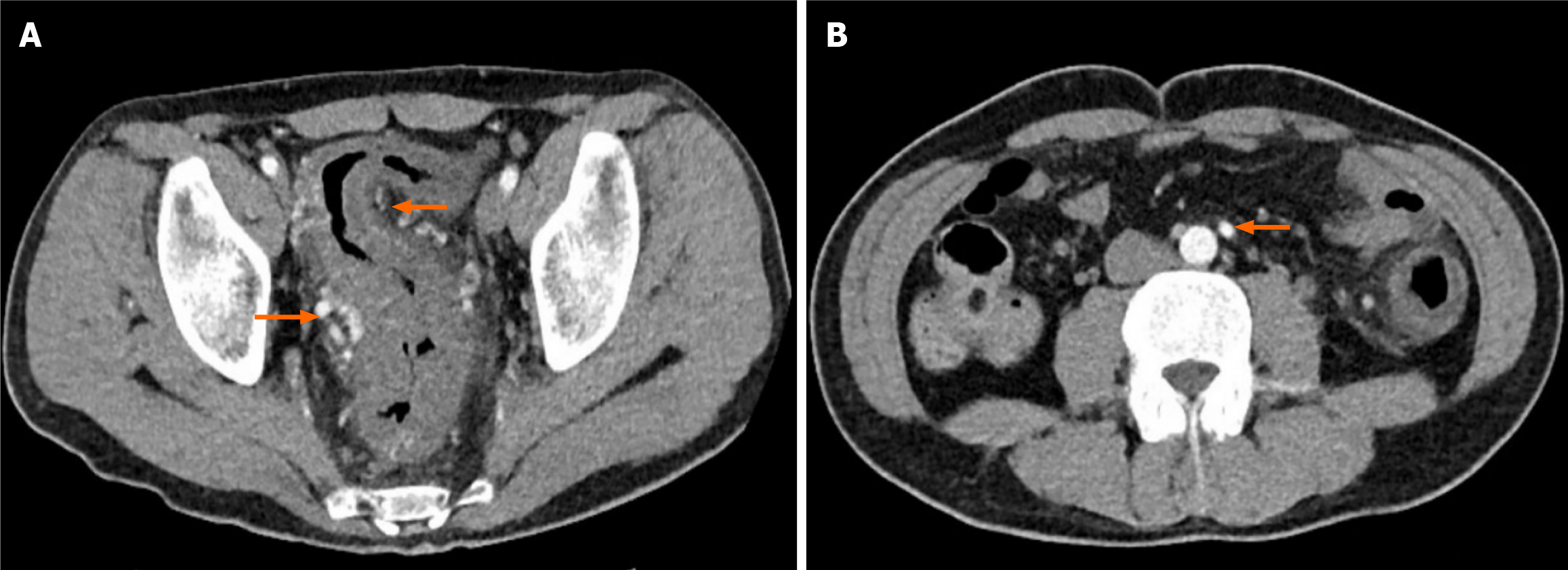
Figure 2 Contrast-enhanced computed tomography.
A: Diffuse thickening of the rectum and sigmoid colon with reduced enhancement, increased surrounding mesenteric exudation, and formation of collateral circulation, as indicated by the arrows; B: Compensatory enlargement of the inferior mesenteric artery, as indicated by the arrow.
- Citation: Jin X, Xu TM, Ruan GC, Zhang XF, Chen XY, Li XQ, Tang H, Xu H, Wang QP, Liu W, Li J. Clinical manifestations and imaging features of inferior mesenteric vein-related colon ischemia. World J Gastroenterol 2026; 32(18): 116229
- URL: https://www.wjgnet.com/1007-9327/full/v32/i18/116229.htm
- DOI: https://dx.doi.org/10.3748/wjg.v32.i18.116229