
















Copyright: ©Author(s) 2026.
World J Gastroenterol. May 14, 2026; 32(18): 116018
Published online May 14, 2026. doi: 10.3748/wjg.v32.i18.116018
Published online May 14, 2026. doi: 10.3748/wjg.v32.i18.116018
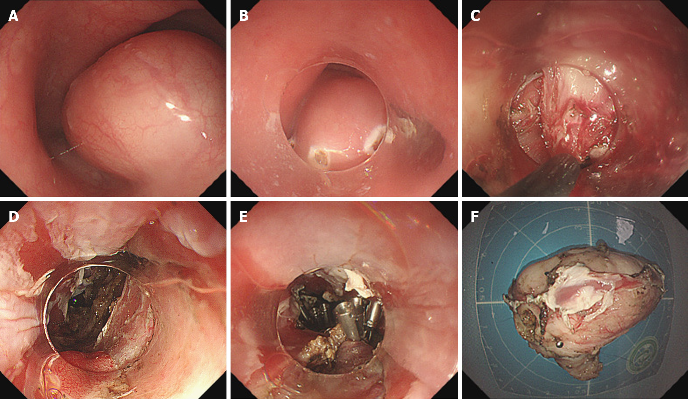
Figure 2 Procedures of endoscopic full-thickness resection for a case with esophageal subepithelial lesion originating from muscularis propria.
A: Endoscopic view of esophageal subepithelial lesion originating from muscularis propria; B: Marking dots placed on the surface of the lesion; C: Dissection of the lesion from the muscularis propria; D: Esophageal wall defect following lesion resection; E: Incomplete closure of the esophageal wall defect using metallic clips; F: Resected specimen.
- Citation: Li XX, Zheng QF, Garcia AM, Shi ZY, Shi M, Islam MS, Liu D. Exposed endoscopic full-thickness resection vs thoracoscopic surgery for complex esophageal subepithelial lesions arising from the muscularis propria. World J Gastroenterol 2026; 32(18): 116018
- URL: https://www.wjgnet.com/1007-9327/full/v32/i18/116018.htm
- DOI: https://dx.doi.org/10.3748/wjg.v32.i18.116018