
















Copyright: ©Author(s) 2026.
World J Gastroenterol. May 7, 2026; 32(17): 119419
Published online May 7, 2026. doi: 10.3748/wjg.v32.i17.119419
Published online May 7, 2026. doi: 10.3748/wjg.v32.i17.119419
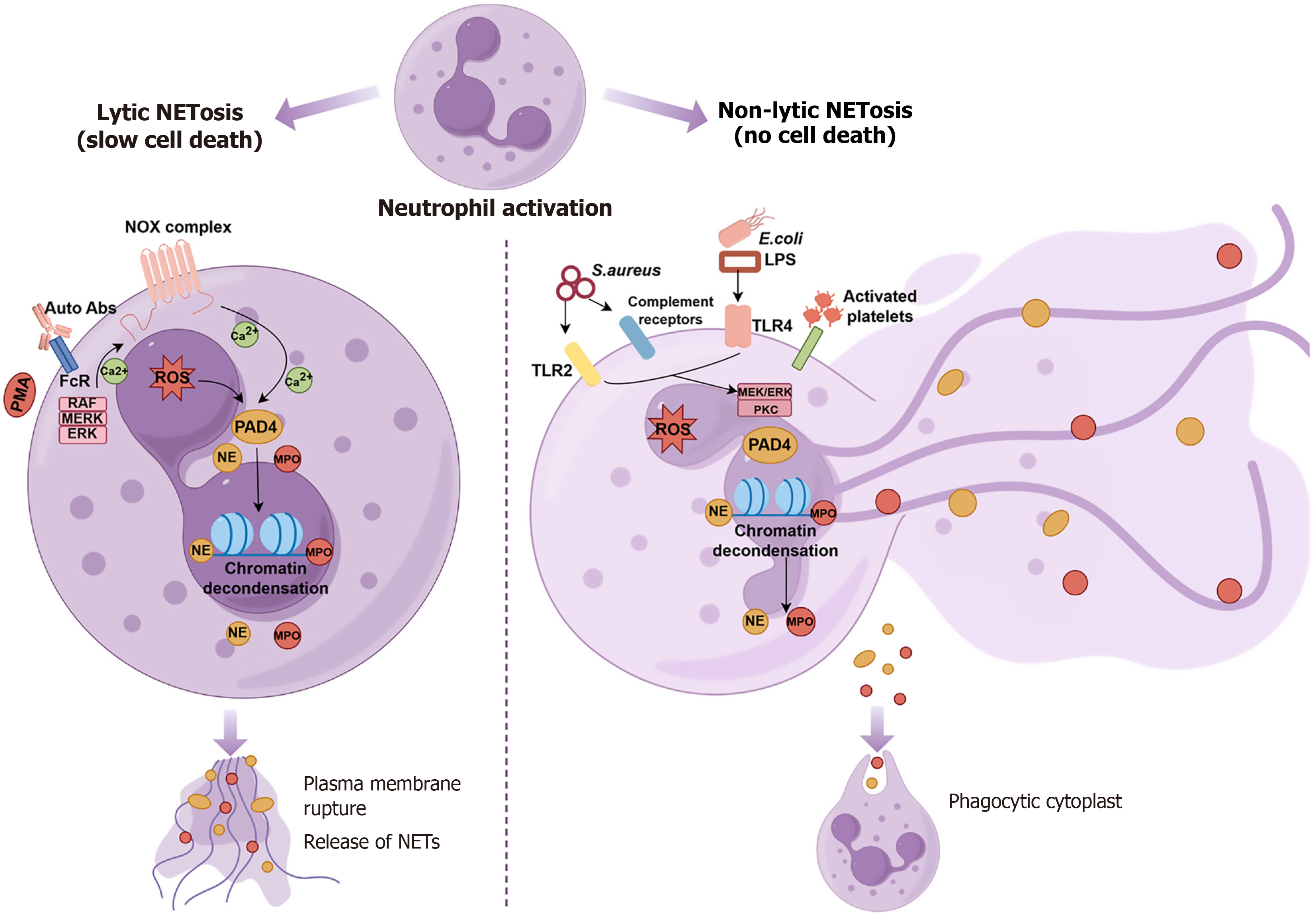
Figure 1 Two main types of neutrophil extracellular traps formation: Suicidal (lytic) and non-suicidal (vital).
Suicidal state of neutrophils with neutrophil extracellular traps formation (NETosis) can be triggered by phorbol myristate acetate, antibodies bounding to the Fc receptor. The Raf-mitogen-activated protein kinase-extracellular regulated protein kinases pathway and nicotinamide adenine dinucleotide phosphate oxidase-dependent reactive oxygen species production are activated. Activation of peptidylarginine deiminase (PAD) 4 results in histone citrullination, followed by exocytosis of decondensed chromatin and enzymes like neutrophil elastase (NE) and myeloperoxidase (MPO). Vital NETosis can be triggered by Staphylococcus aureus infection recognized by Toll-like receptor (TLR) 2 or complement receptors, or by Escherichia coli directly via TLR4 or indirectly via TLR4-activated platelets. Activated PAD4 in turn translocates to the nucleus converting arginine to citrulline on histones, and resulting in chromatin depolymerization, along with its extracellular release including NE and MPO. NOX: Nicotinamide adenine dinucleotide phosphate-oxidase; Abs: Antibodies; FcR: Fc receptor; PMA: Phorbol myristate acetate; MERK: Mitogen-activated protein kinase; ERK: Extracellular regulated protein kinases; Ca2+: Calcium ion; ROS: Reactive oxygen species; PAD4: Peptidylarginine deiminase 4; MPO: Myeloperoxidase; NE: Neutrophil elastase; NETs: Neutrophil extracellular traps; S. aureus: Staphylococcus aureus; E. coli: Escherichia coli; LPS: Lipopolysaccharide; PKC: Protein kinase C; TLR: Toll-like receptor; MEK: Mitogen-activated protein kinase kinase.
- Citation: Qi Y, Ma L, Zhang Y, Liu Y, Su M, Cai TT, Wang M, Sun KW. Insights into the pathogenic roles and targeted therapy of neutrophil extracellular traps in inflammatory bowel disease. World J Gastroenterol 2026; 32(17): 119419
- URL: https://www.wjgnet.com/1007-9327/full/v32/i17/119419.htm
- DOI: https://dx.doi.org/10.3748/wjg.v32.i17.119419