
















Copyright: ©Author(s) 2026.
World J Gastroenterol. May 7, 2026; 32(17): 117479
Published online May 7, 2026. doi: 10.3748/wjg.v32.i17.117479
Published online May 7, 2026. doi: 10.3748/wjg.v32.i17.117479
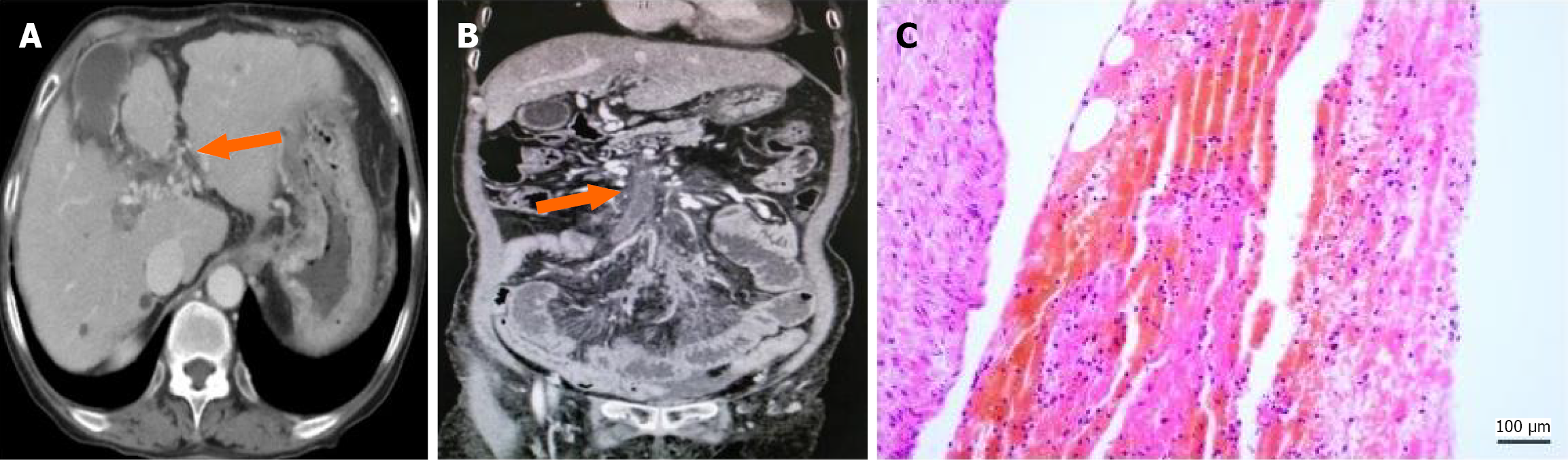
Figure 5 Mixed portal vein thrombosis (cavernous transformation with chronic thrombosis).
A: Contrast-enhanced abdominal computed tomography (CT) (axial view): The proximal main portal vein does not show the normal anatomical structure, with collateral vessels having formed (orange arrow); B: Contrast-enhanced abdominal CT (coronal view): A somewhat low-density vascular shadow on the distal main portal vein and superior mesenteric vein, with relatively homogeneous density consistent with vascular morphology (orange arrow). Increased thickness of the vascular wall with numerous collateral formations in the periphery; C: Histological analysis with hematoxylin and eosin staining: Consistent with the imaging findings, a laminated thrombus is evident microscopically, accompanied by thickening of the vascular wall.
- Citation: Lv YF, Liu BW, Han Y, Meng MM, Li DZ, Tian H, Wang FC, Dong JH, Yang YP, Zhou GD, Ding HG, Zhang YN, Liu FQ, Zhu B. Proposal of a new classification scheme for complete portal vein thrombosis and its clinical significance: A retrospective study. World J Gastroenterol 2026; 32(17): 117479
- URL: https://www.wjgnet.com/1007-9327/full/v32/i17/117479.htm
- DOI: https://dx.doi.org/10.3748/wjg.v32.i17.117479