
















Copyright: ©Author(s) 2026.
World J Gastroenterol. May 7, 2026; 32(17): 117479
Published online May 7, 2026. doi: 10.3748/wjg.v32.i17.117479
Published online May 7, 2026. doi: 10.3748/wjg.v32.i17.117479
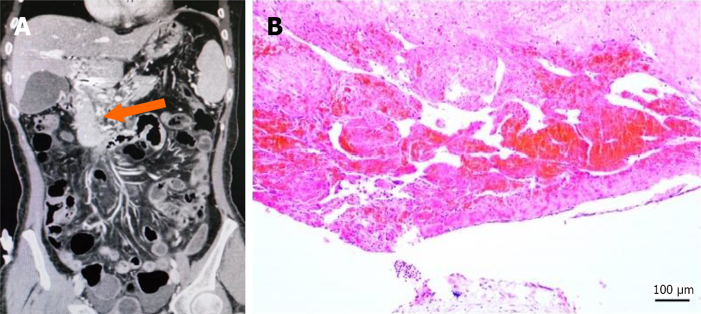
Figure 4 Portal vein cavernous transformation.
A: Contrast-enhanced abdominal computed tomography (coronal view): The complete disappearance of the main portal vein anatomy. Numerous collateral branches had formed at the main portal vein (orange arrow); B: Histological analysis with hematoxylin and eosin staining: Consistent with the imaging findings, the lumen shows aggregates of red blood cells, along with fibrous tissue and prominent proliferation of small vessels (scale bar, 100 μm).
- Citation: Lv YF, Liu BW, Han Y, Meng MM, Li DZ, Tian H, Wang FC, Dong JH, Yang YP, Zhou GD, Ding HG, Zhang YN, Liu FQ, Zhu B. Proposal of a new classification scheme for complete portal vein thrombosis and its clinical significance: A retrospective study. World J Gastroenterol 2026; 32(17): 117479
- URL: https://www.wjgnet.com/1007-9327/full/v32/i17/117479.htm
- DOI: https://dx.doi.org/10.3748/wjg.v32.i17.117479