
















Copyright: ©Author(s) 2026.
World J Gastroenterol. Apr 21, 2026; 32(15): 116679
Published online Apr 21, 2026. doi: 10.3748/wjg.v32.i15.116679
Published online Apr 21, 2026. doi: 10.3748/wjg.v32.i15.116679
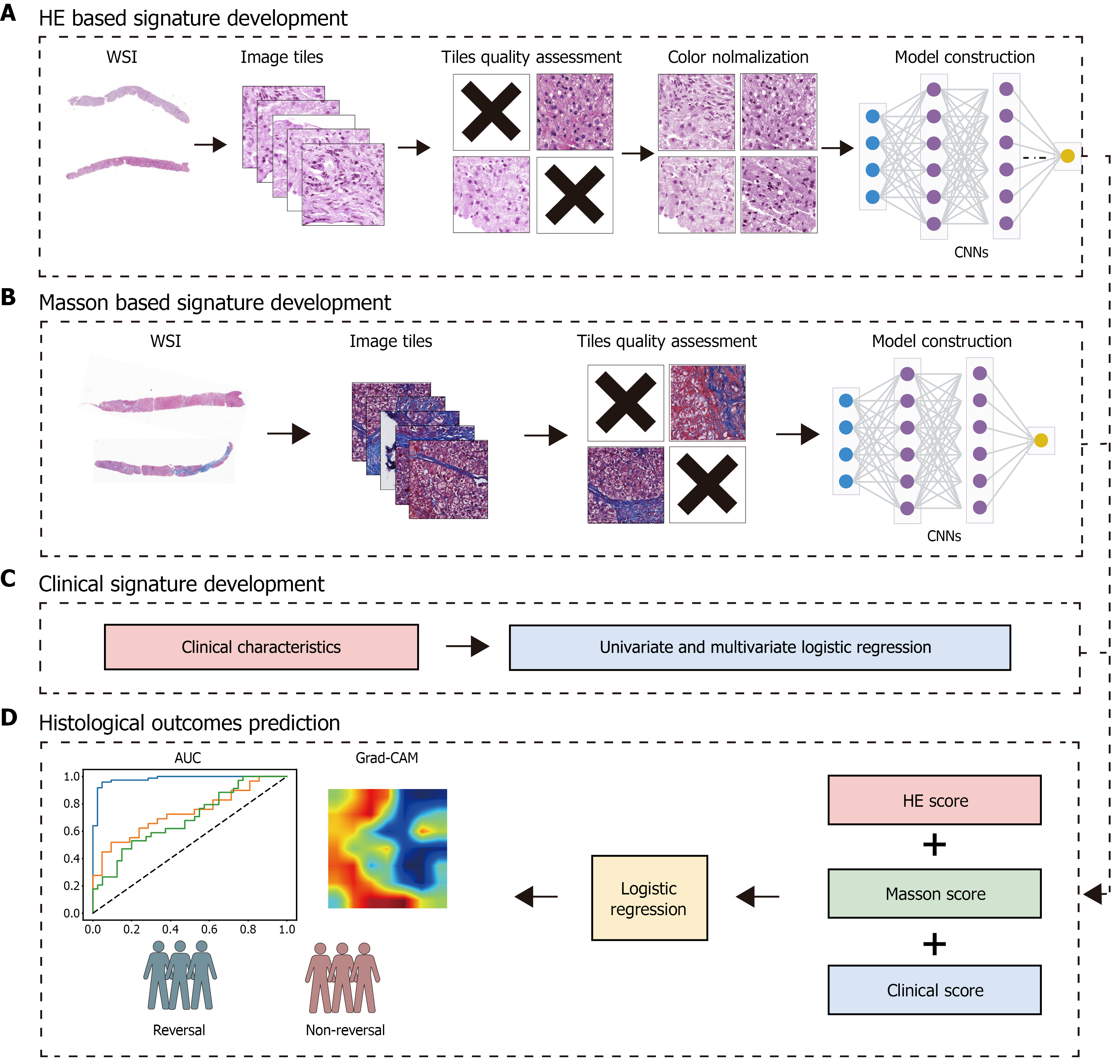
Figure 2 Workflow of the development and testing of deep learning model.
A and B: Digital hematoxylin and eosin (HE)- and Masson-stained slides were first categorized according to whether fibrosis reversal occurred. Whole-slide images were then partitioned into 512 × 512-pixel tiles and subjected to quality control to remove non-informative regions. Color normalization was applied to the HE tiles, whereas Masson-stained tiles were retained in their original color. The quality-controlled tiles, along with their corresponding labels, were used to train convolutional neural networks models; C: Univariate and multivariate logistic regression analyses were performed to identify clinical characteristics associated with histological outcomes; D: A logistic regression fusion model was developed using HE score, Masson score, and clinical score as input variables, followed by performance validation on internal and external validation sets. WSI: Whole-slide image; CNNs: Convolutional neural networks; Grad-CAM: Gradient-weighted Class Activation Mapping; HE: Hematoxylin and eosin; AUC: Area under the receiver operating characteristic curve.
- Citation: Han W, Cheng DY, He QW, Wang SH, Gong SJ, Chen Y, Yang YP. Deep learning-based multimodal model for predicting on-treatment histological outcomes in chronic hepatitis B-associated advanced liver fibrosis. World J Gastroenterol 2026; 32(15): 116679
- URL: https://www.wjgnet.com/1007-9327/full/v32/i15/116679.htm
- DOI: https://dx.doi.org/10.3748/wjg.v32.i15.116679