
















Copyright: ©Author(s) 2026.
World J Gastroenterol. Apr 21, 2026; 32(15): 116364
Published online Apr 21, 2026. doi: 10.3748/wjg.v32.i15.116364
Published online Apr 21, 2026. doi: 10.3748/wjg.v32.i15.116364
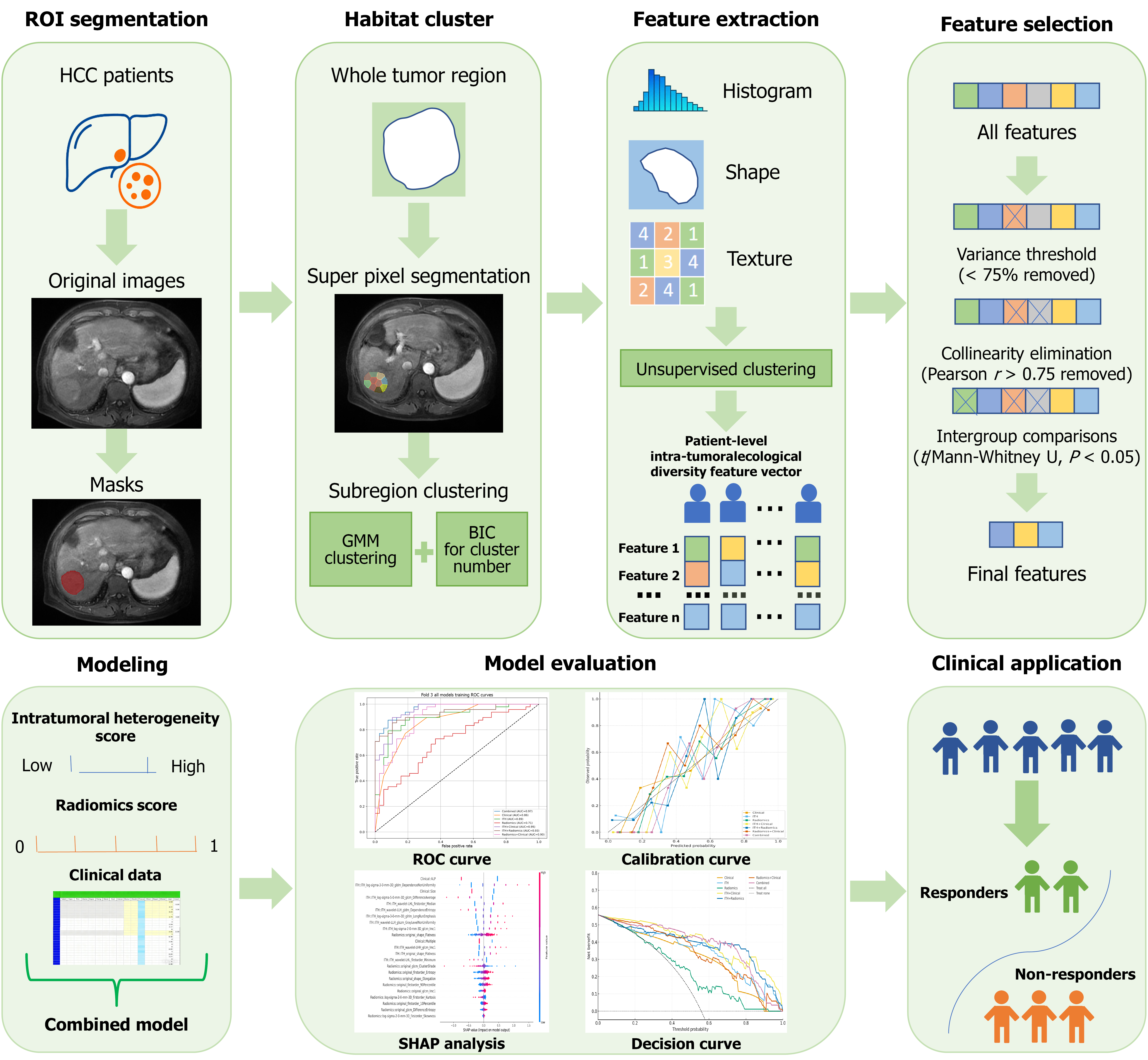
Figure 2 Schematic diagram of the methodological workflow.
The workflow is divided into two parts: The first part is preprocessing and feature acquisition. First, through region of interest segmentation, masks are obtained from the original images of hepatocellular carcinoma patients; then, via habitat clustering, subregion clustering is performed using simple linear iterative clustering superpixels to obtain habitats; subsequently, feature extraction and feature selection are conducted in sequence. The second part is model construction and application: A combined model is built based on the intratumoral heterogeneity score, radiomics score, and clinical data; model evaluation is completed using the receiver operating characteristic curve, calibration curve, SHapley Additive exPlanation analysis, and decision curve; finally, the model is applied in clinical practice to distinguish between responders (treatment responders) and non-responders (non-treatment responders). ROI: Region of interest; HCC: Hepatocellular carcinoma; GMM: Gaussian mixture model; BIC: Bayesian information criterion; ROC: Receiver operating characteristic; SHAP: SHapley Additive exPlanation.
- Citation: Lv JB, Liu W, Wei YG, Tang HN, Chen QQ, Hu HJ, Hu JB. Habitat imaging on contrast-enhanced magnetic resonance imaging predicts early response to transarterial chemoembolization in hepatocellular carcinoma. World J Gastroenterol 2026; 32(15): 116364
- URL: https://www.wjgnet.com/1007-9327/full/v32/i15/116364.htm
- DOI: https://dx.doi.org/10.3748/wjg.v32.i15.116364