
















Copyright: ©Author(s) 2026.
World J Gastroenterol. Apr 21, 2026; 32(15): 115263
Published online Apr 21, 2026. doi: 10.3748/wjg.v32.i15.115263
Published online Apr 21, 2026. doi: 10.3748/wjg.v32.i15.115263
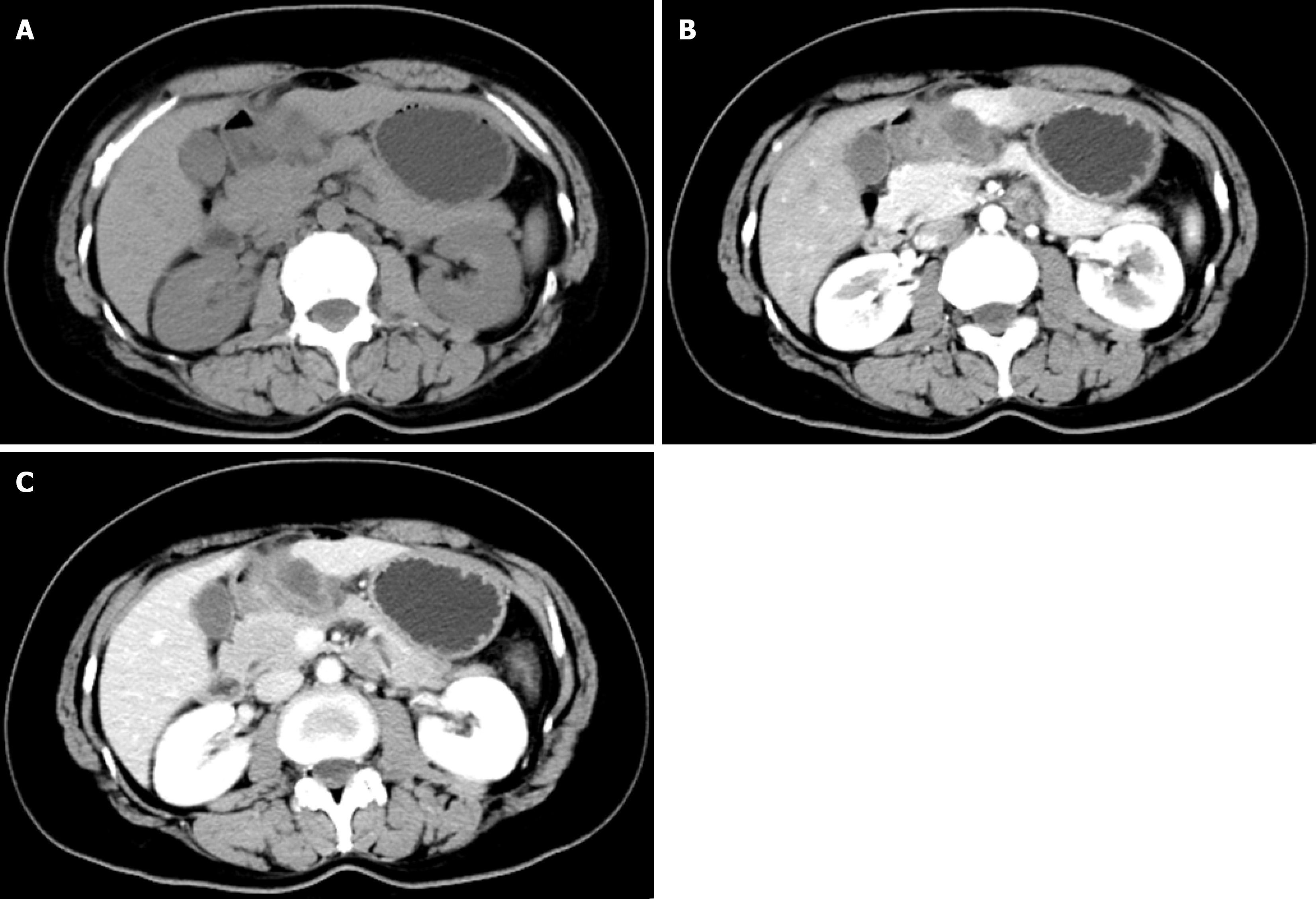
Figure 1 On computed tomography scanning, a cystic lesion is observed outside the lesser curvature wall of the gastric antrum, measuring approximately 3.
0 cm × 2.2 cm. A: After contrast enhancement, mild enhancement is noted at the periphery of the lesion, represents the non-contrast computed tomography (CT) scan phase; B: Represents the arterial phase of the CT scan; C: Represents the venous phase of the CT scan. The lesion exhibits thick-walled changes with blurred margins.
- Citation: Wang JJ, Xu WL, Li AQ. Aggressive angiomyxoma occurring in the stomach: A case report and review of literature. World J Gastroenterol 2026; 32(15): 115263
- URL: https://www.wjgnet.com/1007-9327/full/v32/i15/115263.htm
- DOI: https://dx.doi.org/10.3748/wjg.v32.i15.115263