
















Copyright: ©Author(s) 2026.
World J Gastroenterol. Apr 14, 2026; 32(14): 111455
Published online Apr 14, 2026. doi: 10.3748/wjg.v32.i14.111455
Published online Apr 14, 2026. doi: 10.3748/wjg.v32.i14.111455
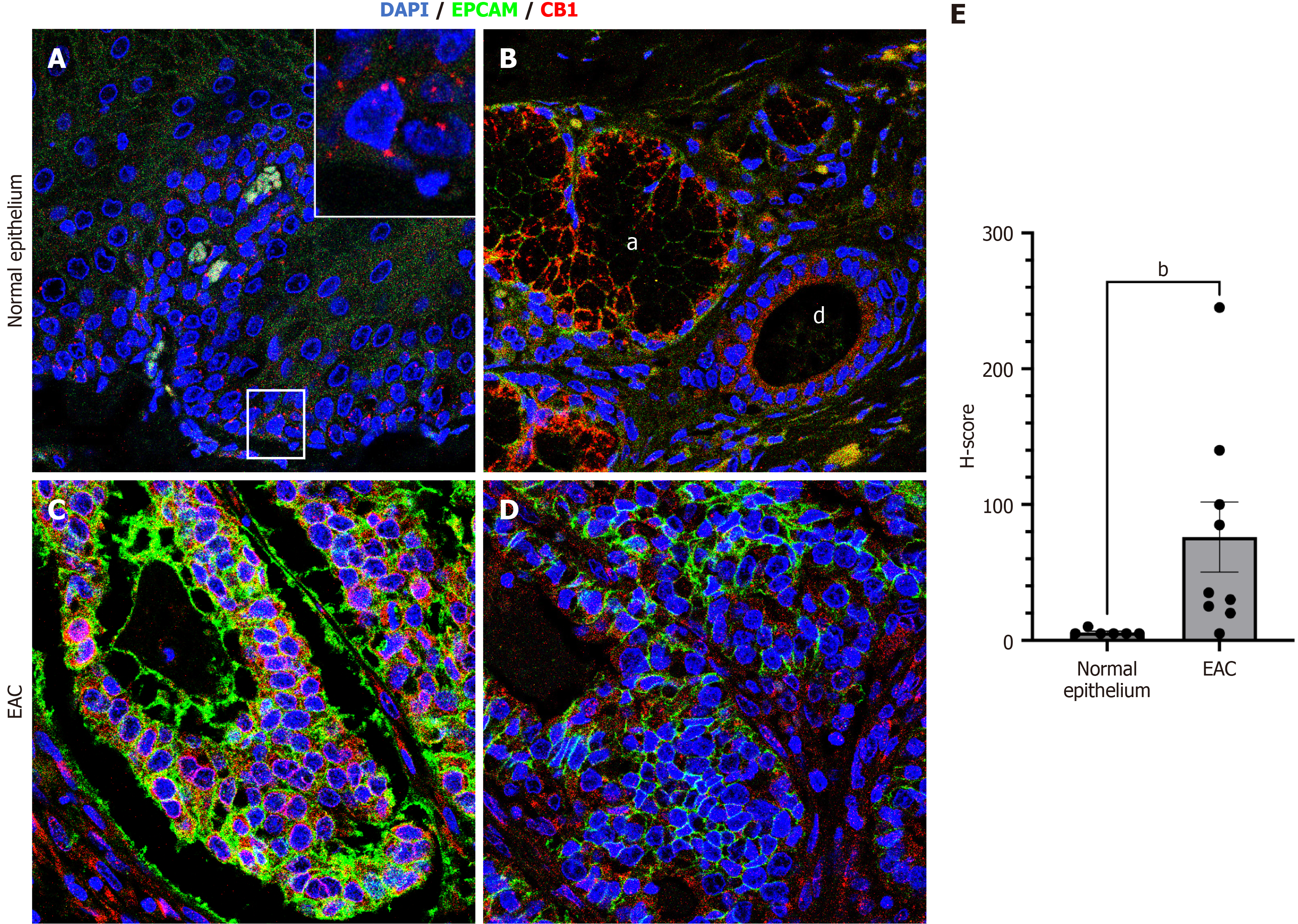
Figure 5 Expression of CB1 in human normal esophageal mucosa and esophageal adenocarcinoma assessed by confocal microscopy.
A: A representative immunofluorescence analysis of CB1 (red) and epithelial cell adhesion molecule (EPCAM) (green) expression in the normal human esophageal mucosa (magnification 63 ×). The inset highlights the CB1 expression in the basal epithelial layer; B: A representative immunofluorescence analysis depicting CB1 and EPCAM co-expression in the ductal epithelium of the submucosal gland (d) and acini of the submucosal gland (a); C and D: Representative immunofluorescence analyses of CB1 and EPCAM co-expression in two distinct human esophageal adenocarcinoma (EAC) samples; E: Bar graph represents a comparative H-score semiquantitative intensity analysis of CB1 expression in human normal esophageal mucosa and EAC. The data were analyzed using the Mann-Whitney U test. bP < 0.01. DAPI: 4’,6-diamidino-2-phenylindole; EPCAM: Epithelial cell adhesion molecule; EAC: Esophageal adenocarcinoma.
- Citation: Goldman A, Gonzalez G, Karpova SA, Buon L, Shammas MA, Mashimo H, Frank MH, Frank NY. Optimal cannabinoid-terpene combination ratios suppress mutagenicity of gastric reflux in normal and metaplastic esophageal cells. World J Gastroenterol 2026; 32(14): 111455
- URL: https://www.wjgnet.com/1007-9327/full/v32/i14/111455.htm
- DOI: https://dx.doi.org/10.3748/wjg.v32.i14.111455