
















Copyright: ©Author(s) 2026.
World J Gastroenterol. Apr 7, 2026; 32(13): 114563
Published online Apr 7, 2026. doi: 10.3748/wjg.v32.i13.114563
Published online Apr 7, 2026. doi: 10.3748/wjg.v32.i13.114563
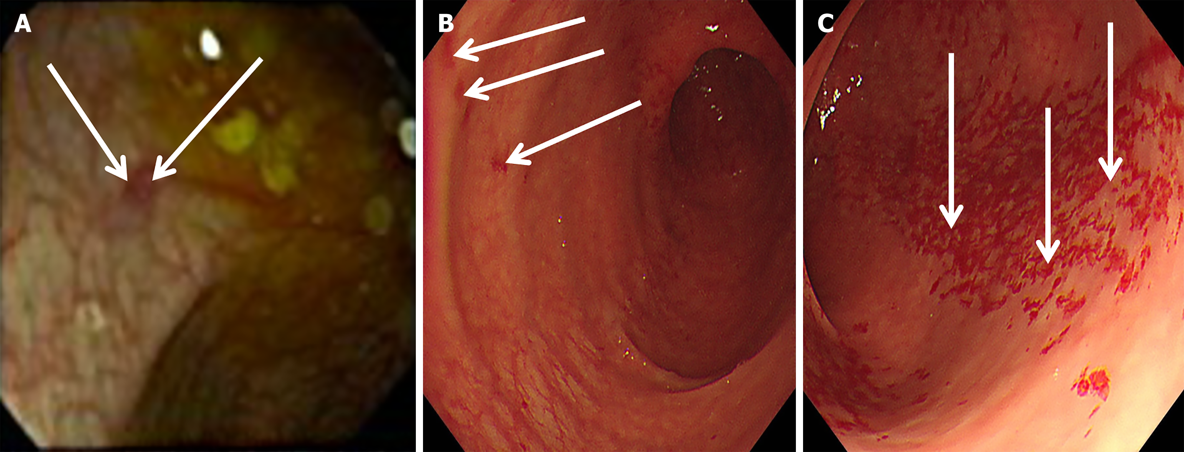
Figure 1 Electronic colonoscopy images on two occasions.
A: On day 8 of diarrhea, electronic colonoscopy (arrows) revealed a patchy erosion measuring approximately 4 mm × 3 mm in the transverse colon (hepatic flexure) (obtained by an experienced endoscopist). Image resolution is limited solely due to suboptimal bowel preparation; however, the colonic erosion remains discernible. The location of the erosion corresponded to the presentation of right upper quadrant pain; B and C: On day 64 of diarrhea, multiple erosions were observed in the ascending colon (arrows in B) and the transverse colon (arrows in C). Compared to panel A, the area of colonic mucosal erosion shown in panels B and C had significantly enlarged, which was consistent with the worsening diarrhea symptoms.
- Citation: Guo T, Wang X, Wang SY, Lin TF. Isolated colonic aspergillosis in an immunocompetent individual diagnosed by metagenomic next-generation sequencing: A case report. World J Gastroenterol 2026; 32(13): 114563
- URL: https://www.wjgnet.com/1007-9327/full/v32/i13/114563.htm
- DOI: https://dx.doi.org/10.3748/wjg.v32.i13.114563