
















Copyright: ©Author(s) 2026.
World J Gastroenterol. Mar 28, 2026; 32(12): 119002
Published online Mar 28, 2026. doi: 10.3748/wjg.v32.i12.119002
Published online Mar 28, 2026. doi: 10.3748/wjg.v32.i12.119002
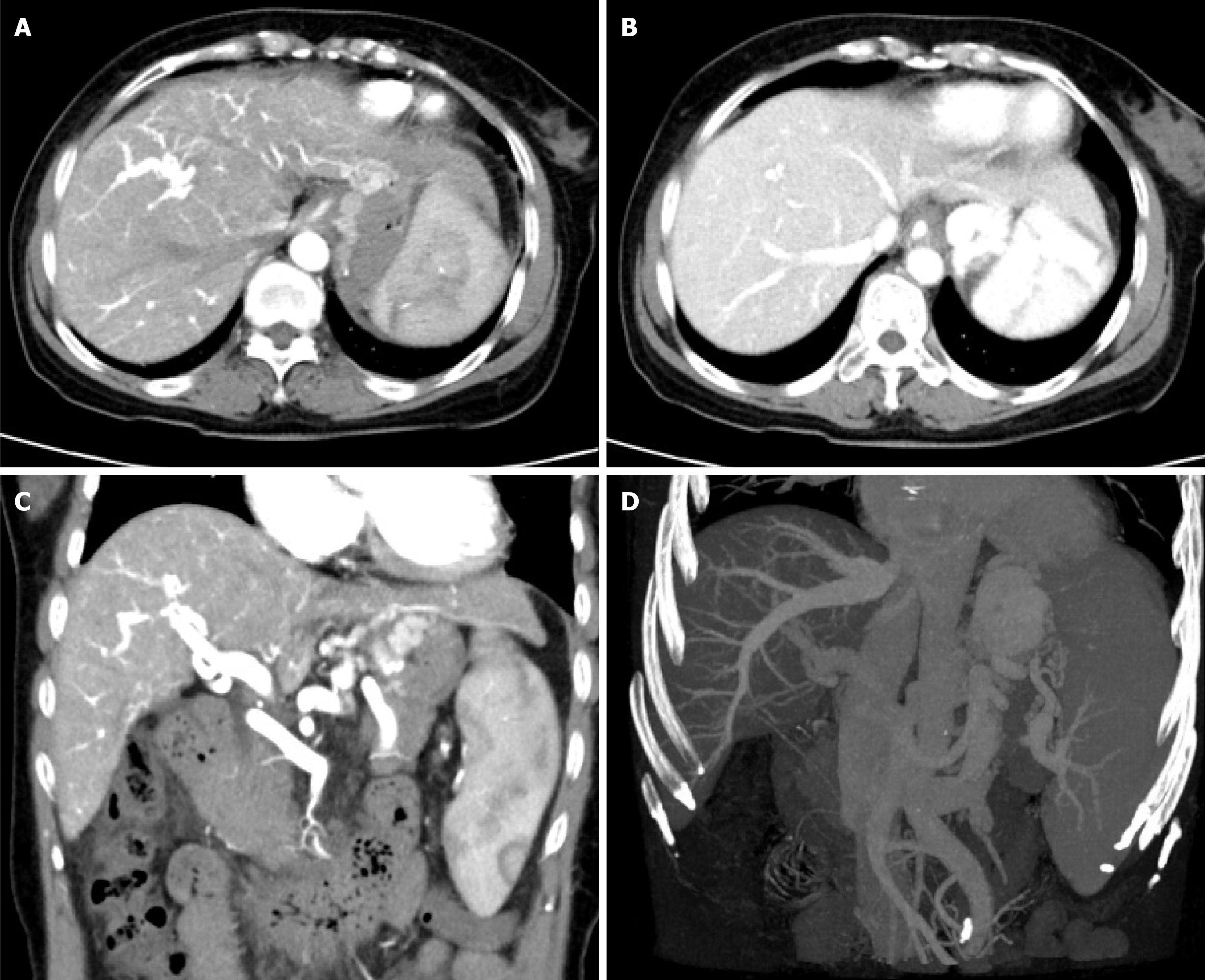
Figure 2 Contrast-enhanced computed tomography.
A: Liver cirrhosis and splenomegaly, arterial phase imaging of the main and left/right branches of the portal vein; B: Small low-density shadows at the edge of the main portal vein and diameter of portal vein 1.5 cm; C and D: Left branch portal vein narrowed with spongiform degeneration and multiple collateral circulation formation.
- Citation: Zhang TQ, Zhang L, Yong X, Tian C, Chen BJ, Qin JP, Mu D, Tang SH. Transjugular intrahepatic portosystemic shunt for variceal bleeding due to hereditary hemorrhagic telangiectasia with cirrhosis: A case report. World J Gastroenterol 2026; 32(12): 119002
- URL: https://www.wjgnet.com/1007-9327/full/v32/i12/119002.htm
- DOI: https://dx.doi.org/10.3748/wjg.v32.i12.119002