
















Copyright: ©Author(s) 2026.
World J Gastroenterol. Mar 21, 2026; 32(11): 115809
Published online Mar 21, 2026. doi: 10.3748/wjg.v32.i11.115809
Published online Mar 21, 2026. doi: 10.3748/wjg.v32.i11.115809
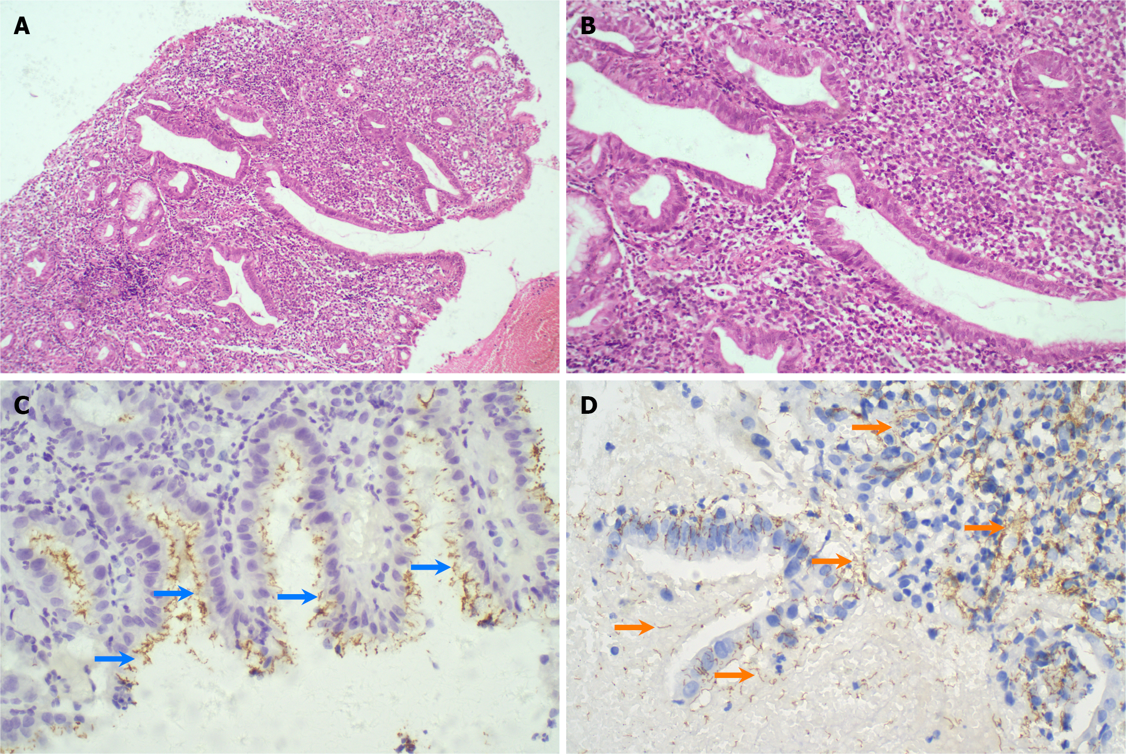
Figure 3 Hematoxylin-eosin staining and immunohistochemistry staining of biopsy tissues.
A: Gastric mucosa revealed severe inflammation [Hematoxylin-eosin (HE) staining, 100 ×]; B: Mainly lymphocyte and plasma cell infiltration (HE staining, 400 ×); C: Helicobacter pylori immunohistochemistry (IHC) staining showing pathogens on the superficial mucosa (IHC staining, 400 ×, blue arrows); D: IHC staining of Treponema pallidum showed thread-like pathogens distributed in the tissue and mucus (IHC staining, 400 ×, orange arrows).
- Citation: Zhang JL, Wei N, Chen TM, Qiu W. Triple infection of gastric syphilis, Helicobacter pylori and human immunodeficiency virus: A case report. World J Gastroenterol 2026; 32(11): 115809
- URL: https://www.wjgnet.com/1007-9327/full/v32/i11/115809.htm
- DOI: https://dx.doi.org/10.3748/wjg.v32.i11.115809