
















Copyright: ©Author(s) 2026.
World J Gastroenterol. Mar 21, 2026; 32(11): 115393
Published online Mar 21, 2026. doi: 10.3748/wjg.v32.i11.115393
Published online Mar 21, 2026. doi: 10.3748/wjg.v32.i11.115393
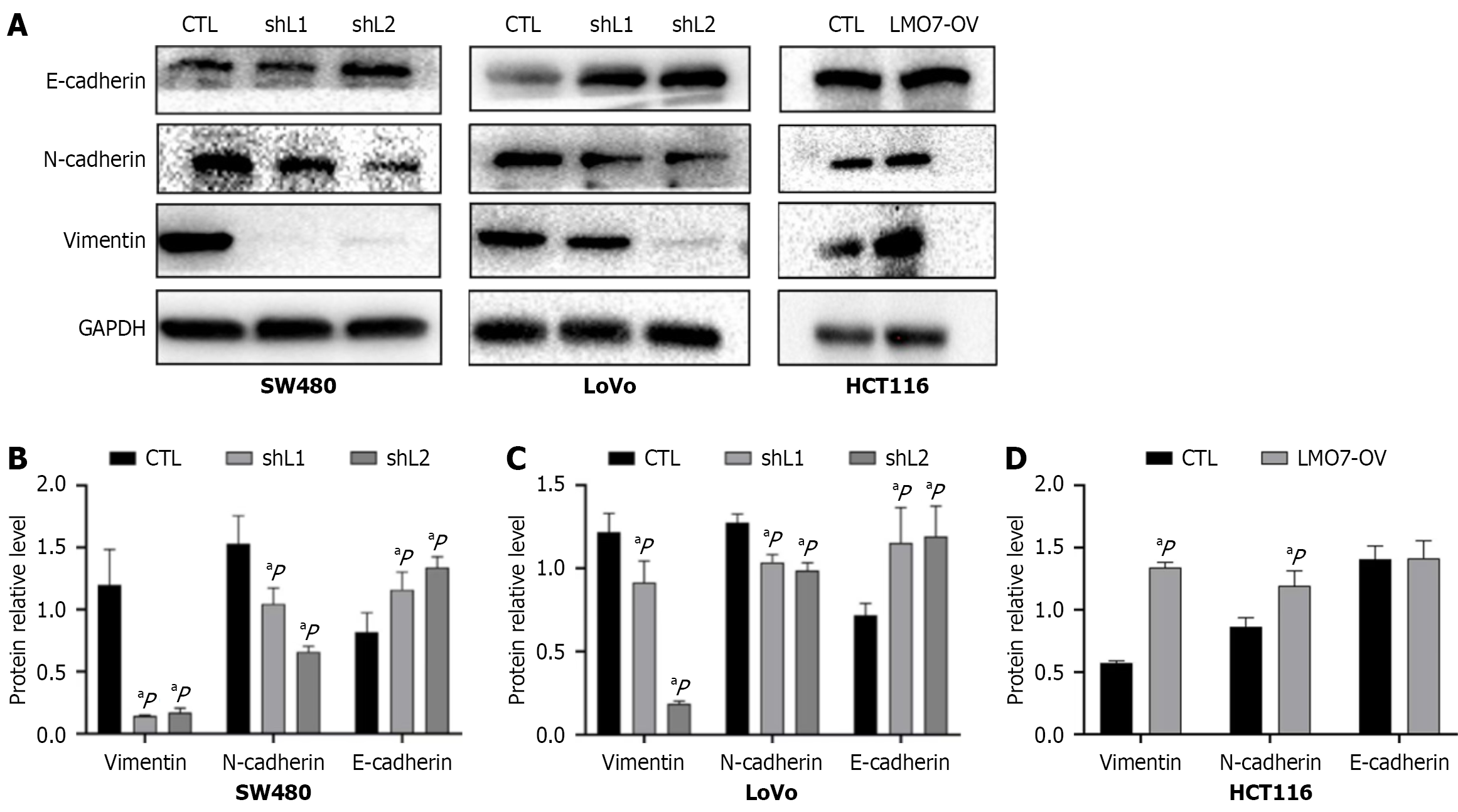
Figure 5 Expression of the E-cadherin, N-cadherin, and vimentin proteins was detected by Western blotting.
A: Western blotting; B: Statistical graph for SW480 cells; C: Statistical graph for LoVo cells; D: Statistical graph for HCT116 cells. Using the Kruskal-Wallis. aP < 0.05. CTL: Cells transfected with the black vector; GAPDH: Glyceraldehyde-3-phosphate dehydrogenase; LMO7-OV: Cells overexpressing LIM domain only protein 7; shL1: Cells transfected with sh-LMO7-1; shL2: Cells transfected with sh-LMO7-2.
- Citation: Bai X, Zheng B, Li XM, Wang JD, Huang XH, Fu SM, Chen X. LIM domain only protein 7 promotes metastasis in colorectal cancer by TGF-β/ZEB1 pathway. World J Gastroenterol 2026; 32(11): 115393
- URL: https://www.wjgnet.com/1007-9327/full/v32/i11/115393.htm
- DOI: https://dx.doi.org/10.3748/wjg.v32.i11.115393