











BPG is committed to discovery and dissemination of knowledge
Retrospective Study
©Author(s) (or their employer(s)) 2026.
World J Gastroenterol. Mar 14, 2026; 32(10): 116152
Published online Mar 14, 2026. doi: 10.3748/wjg.v32.i10.116152
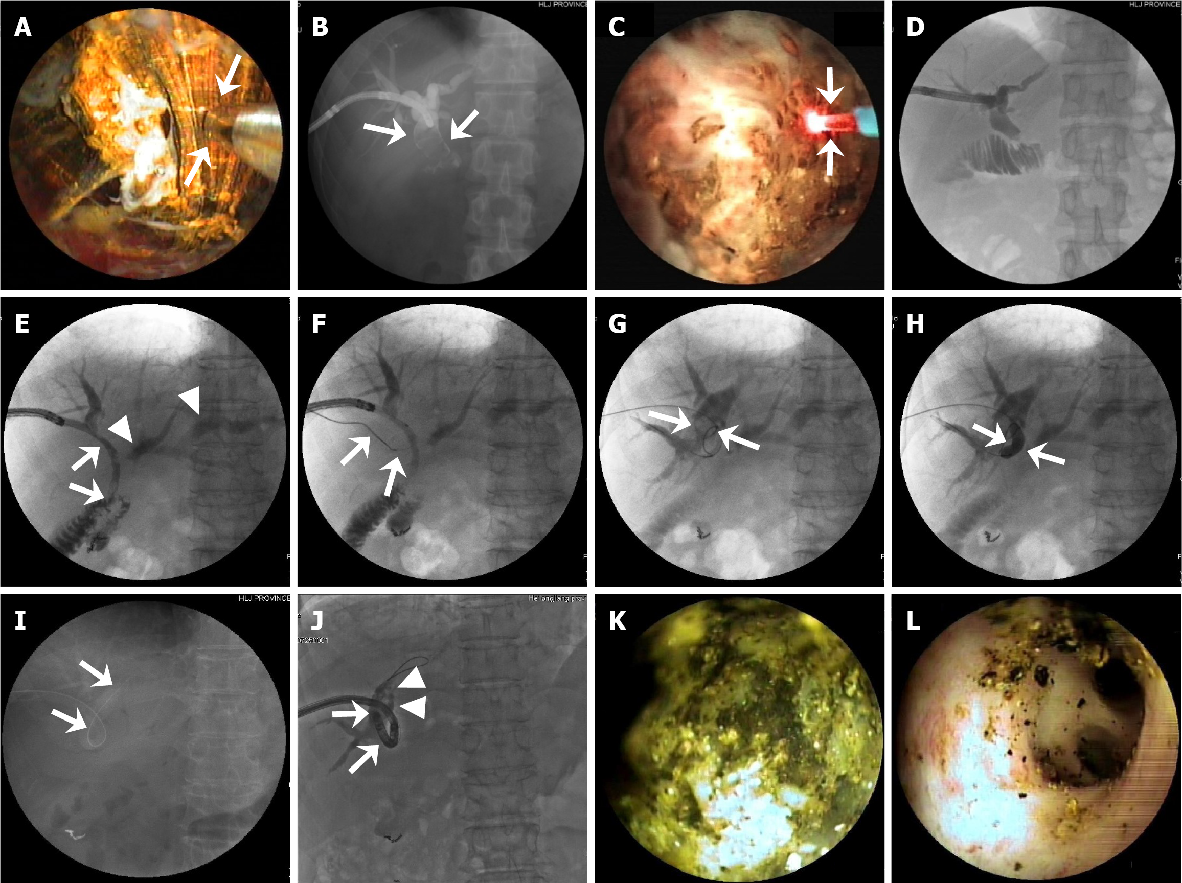
Figure 4
Figure 4 Representative images of successful percutaneous transhepatic cholangioscopy-assisted biliary stone therapy with electrohydraulic lithotripsy and laser lithotripsy for biliary impacted stones, and intrahepatic stones. A: In a patient with a large stone in intrahepatic duct, percutaneous transhepatic cholangioscopy (PTCS) shows a stone being treated with electrohydraulic lithotripsy (EHL) (arrow points to EHL probe); B-D: In a patient with stones impacted in the extra-hepatic bile duct (arrow), percutaneous transhepatic cholangiography (PTC) shows stones larger than 2 cm (B). PTCS shows a stone with laser lithotripsy (LL) (arrow points to the laser probe) (C), and PTC reveals complete stone clearance (D) after PTCS-assisted LL; E-L: In a patient with a complex stricture and bilateral stones, after the removal of the stones in the left and right hepatic duct, PTCS shows the right hepatic duct and contrast medium passing through the mild anastomotic stricture (arrows), and the dilated left hepatic duct and branch bile duct (arrowheads) with the strictures, but fails to show the right posterior branch duct or stones in the duct (E). Under the guidance of PTCS, a guidewire enters the right posterior branch duct (arrows) (F). PTC shows a 1.4 cm × 1.9 cm stone in the right posterior upper branch duct (G). After successful balloon dilation of the stricture (arrows) (H), an indwelling PTCS catheter is placed and left in the duct (I) for 1 week. A cholangioscope was inserted into the right posterior upper branch duct (J), and shows multiple stones (arrows) (K). Under PTCS guidance, the stones were successfully removed with a basket mechanical lithotripsy, and washed out of the duct (L).


Write to the Help Desk