
















©Author(s) (or their employer(s)) 2026.
World J Gastroenterol. Mar 14, 2026; 32(10): 116152
Published online Mar 14, 2026. doi: 10.3748/wjg.v32.i10.116152
Published online Mar 14, 2026. doi: 10.3748/wjg.v32.i10.116152
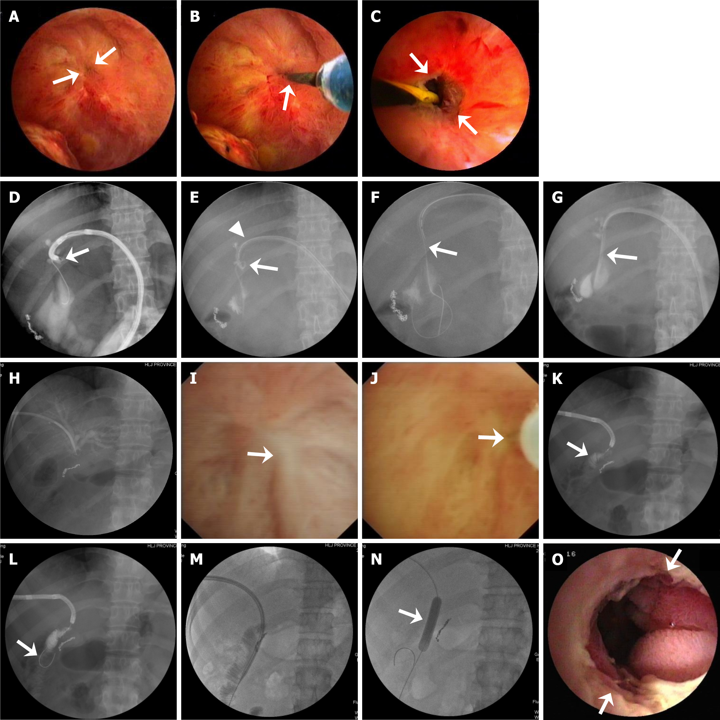
Figure 3 Representative images showing successful percutaneous transhepatic cholangioscopy-assisted anastomotic recanalization in patients with severe benign bilioenteric anastomotic stricture with a guidewire, the microcatheter system, and a needle knife.
A-C: In a patient with a small dimple anastomotic site (arrow) in the center of a fibrous scar (converging mucosal membrane) (A), a zebra guidewire (via an endoscopic retrograde cholangiopancreatography cannula) with a flexible tip was used to tap the mucosa (arrow) in the site of the dimple (B) and after the guidewire passed through the stenotic anastomosis, initial dilation (arrow points to the open anastomosis) for the stricture was performed with a fine bougie (C); D-G: In a patient with a severe anastomotic stricture, a stiff 0.025-inch micro-guidewire, guided under percutaneous transhepatic cholangioscopy (PTCS), was looped through stenotic anastomosis of sharp angulation and looping (arrow in D). Then a 5-Fr microcatheter (≤ 3 Fr at the tip) was looped (arrow) to pass through the stricture and kept in place within the PTCS catheter (arrowhead points to the tip of the PTCS catheter above the stricture) (E). The looping stricture was straightened with a dreamwire (arrow) under fluoroscopy one month later (F), and an 18-Fr PTCS catheter (arrow) was finally placed through the stricture after stepwise bougie dilation (G); H-O: In a patient with complete bilioenteric anastomotic membranous occlusion, percutaneous transhepatic cholangiography shows no passage of the contrast medium through the anastomosis (H). PTCS only shows the converging mucosal membrane (arrow) in the area of scars, but not the anastomosis (I). A 25-gauge injection needle (arrow) was used to puncture the center of the scars under direct visualization (J). Radiography shows the jejunal circular folds (arrow) (K). After a small incision was made with a needle knife at the puncture site, a guidewire was inserted, which was confirmed in the jejunum (arrow) (L). Then, the stenotic anastomosis was dilated with a bougie (M) and a 10-mm balloon with no remaining waist (arrow) (N). PTCS shows enlargement of the anastomosis (arrows) after balloon dilation for nine days (O), indicating successful recanalization and dilation.
- Citation: Ren X, Wang C, Qu YP, Tang XF, Xia T, Lu YX, Sun XM. Percutaneous transhepatic cholangioscopy-assisted procedures for the management of postoperative benign bilioenteric anastomotic strictures with or without biliary stones. World J Gastroenterol 2026; 32(10): 116152
- URL: https://www.wjgnet.com/1007-9327/full/v32/i10/116152.htm
- DOI: https://dx.doi.org/10.3748/wjg.v32.i10.116152