
















©Author(s) (or their employer(s)) 2026.
World J Gastroenterol. Mar 14, 2026; 32(10): 113771
Published online Mar 14, 2026. doi: 10.3748/wjg.v32.i10.113771
Published online Mar 14, 2026. doi: 10.3748/wjg.v32.i10.113771
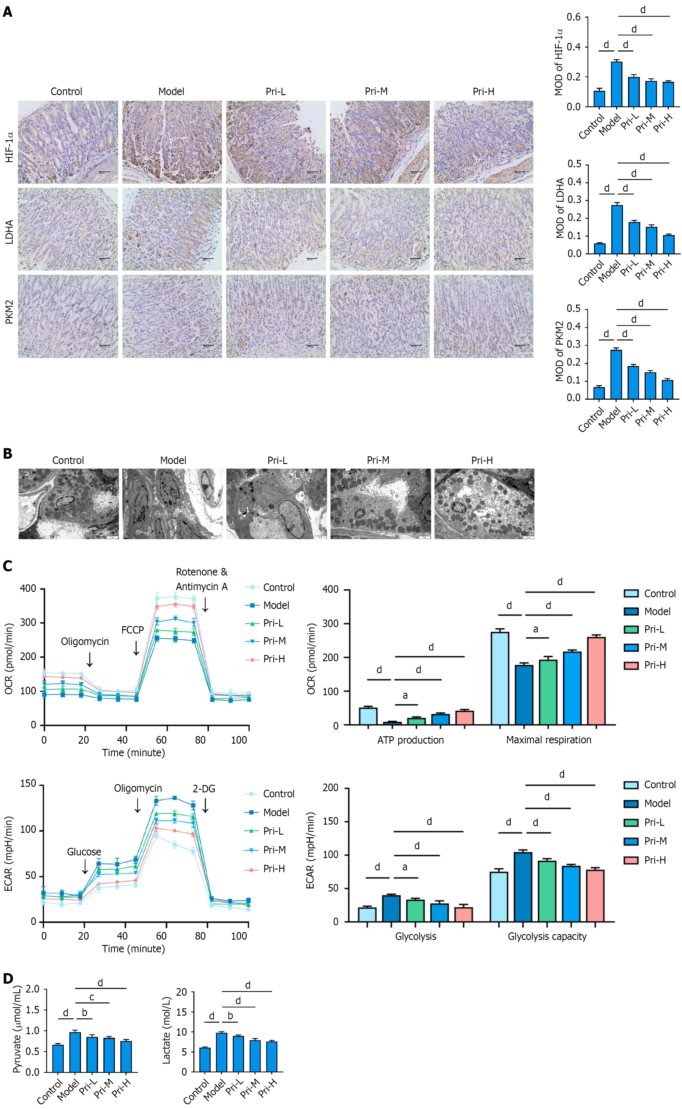
Figure 4 Pristimerin regulates glycolytic reprogramming in gastric mucosal cells.
A: Inhibitory effects of pristimerin on key glycolytic regulators (hypoxia-inducible factor-1α, lactate dehydrogenase A, pyruvate kinase M2) in the gastric mucosa of mice. Scale bar, 100 μm; B: Representative transmission electron microscopy images revealing ultrastructural features in mouse gastric mucosal cells. Scale bar, 2 μm; C: Analysis of cellular energy metabolism by Seahorse XF technology in human gastric epithelial (GES-1) cells following treatment with pristimerin; D: Quantitative analysis of pyruvate and lactate production in GES-1 cells. aP < 0.05. bP < 0.01. cP < 0.001. dP < 0.0001. Values represent the mean ± SD (A: n = 6; C and D: n = 5). HIF-1α: Hypoxia-inducible factor-1α; LDHA: Lactate dehydrogenase A; PKM2: Pyruvate kinase M2; Pri-L: Low-dose pristimerin; Pri-M: Medium-dose pristimerin; Pri-H: High-dose pristimerin; MOD: Mean optical density; FCCP: Trifluoromethoxy carbonylcyanide phenylhydrazone; 2-DG: 2-deoxy-D-glucose; OCR: Oxygen consumption rate; ECAR: Extracellular acidification rate.
- Citation: Wen JS, Pan ZW, Yao XD, Liu YQ, Zhu YD. Pristimerin ameliorates spasmolytic polypeptide-expressing metaplasia by modulating Cdkn1c (p57)-mediated glycolytic reprogramming. World J Gastroenterol 2026; 32(10): 113771
- URL: https://www.wjgnet.com/1007-9327/full/v32/i10/113771.htm
- DOI: https://dx.doi.org/10.3748/wjg.v32.i10.113771