
















©The Author(s) 2026.
World J Gastroenterol. Jan 7, 2026; 32(1): 112090
Published online Jan 7, 2026. doi: 10.3748/wjg.v32.i1.112090
Published online Jan 7, 2026. doi: 10.3748/wjg.v32.i1.112090
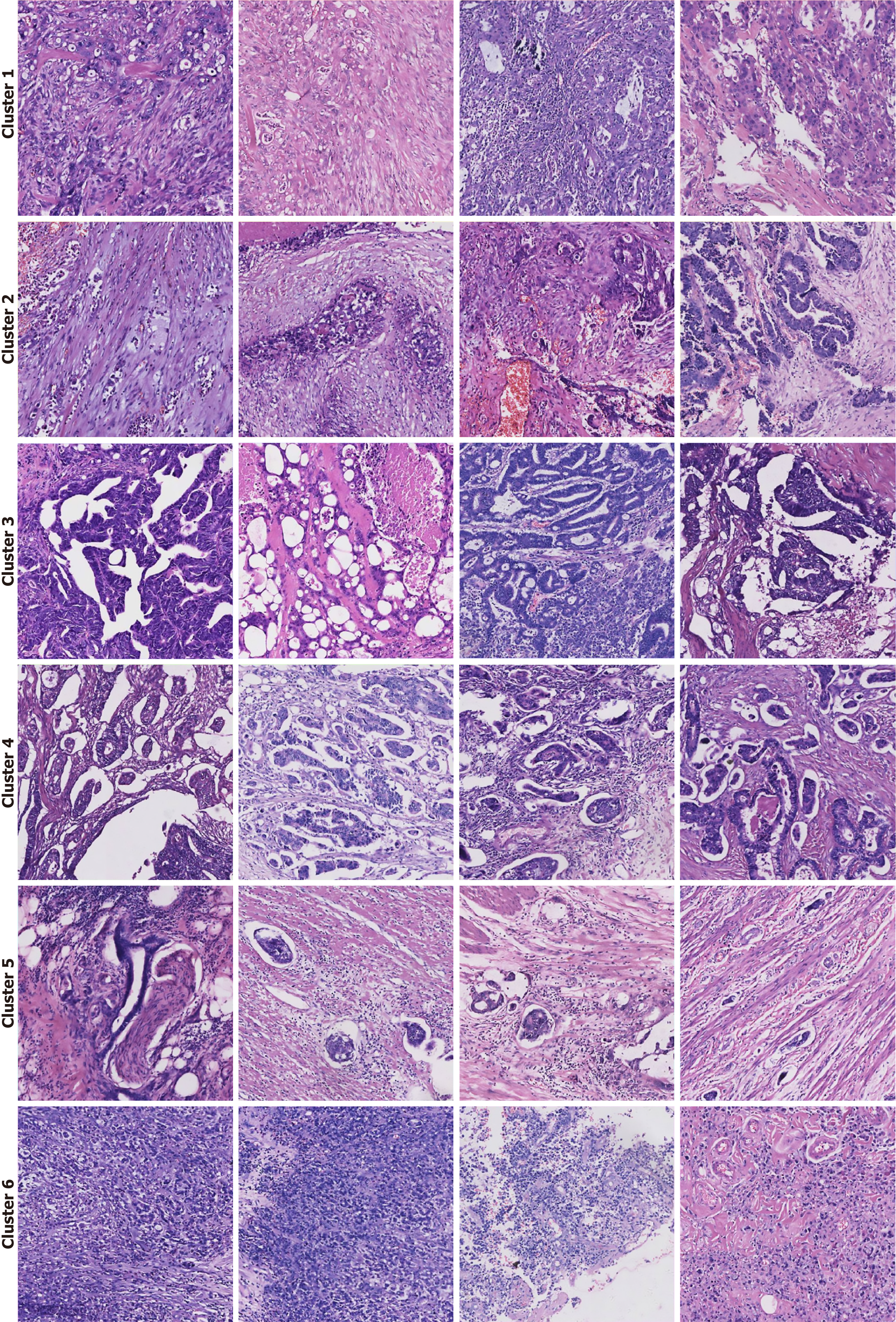
Figure 7 Histopathological interpretation of high-attention morphological clusters.
Representative image tiles from the six distinct clusters identified by the deep learning model. Each cluster corresponds to a specific histopathological phenotype associated with lymph node metastasis risk. Cluster 1: Poorly differentiated adenocarcinoma, characterized by solid sheets and trabecular growth patterns with a high nuclear-to-cytoplasmic ratio. Cluster 2: Prominent desmoplastic reaction, showing a dense fibroblastic stromal response to invading tumor cells. Cluster 3: Adenocarcinoma with complex glandular architecture, featuring cribriform, fused, or back-to-back glands indicative of high-grade tumor organization. Cluster 4: Micropapillary adenocarcinoma, a high-risk pattern defined by small, cohesive tufts of tumor cells floating in stromal spaces without a true fibrovascular core. Cluster 5: Overt invasion, showcasing clear evidence of lymphovascular invasion and perineural invasion, where tumor cells infiltrate lymphatic channels and surround nerve fibers. Cluster 6: Signet-ring cell carcinoma, a distinct subtype composed of tumor cells containing large intracytoplasmic mucin vacuoles that displace the nucleus to the periphery.
- Citation: Zou LF, Wang XB, Li JW, Ouyang X, Luo YY, Luo Y, Wang CL. Predicting lymph node metastasis in colorectal cancer using case-level multiple instance learning. World J Gastroenterol 2026; 32(1): 112090
- URL: https://www.wjgnet.com/1007-9327/full/v32/i1/112090.htm
- DOI: https://dx.doi.org/10.3748/wjg.v32.i1.112090