
















©The Author(s) 2025.
World J Gastroenterol. Feb 28, 2025; 31(8): 100146
Published online Feb 28, 2025. doi: 10.3748/wjg.v31.i8.100146
Published online Feb 28, 2025. doi: 10.3748/wjg.v31.i8.100146
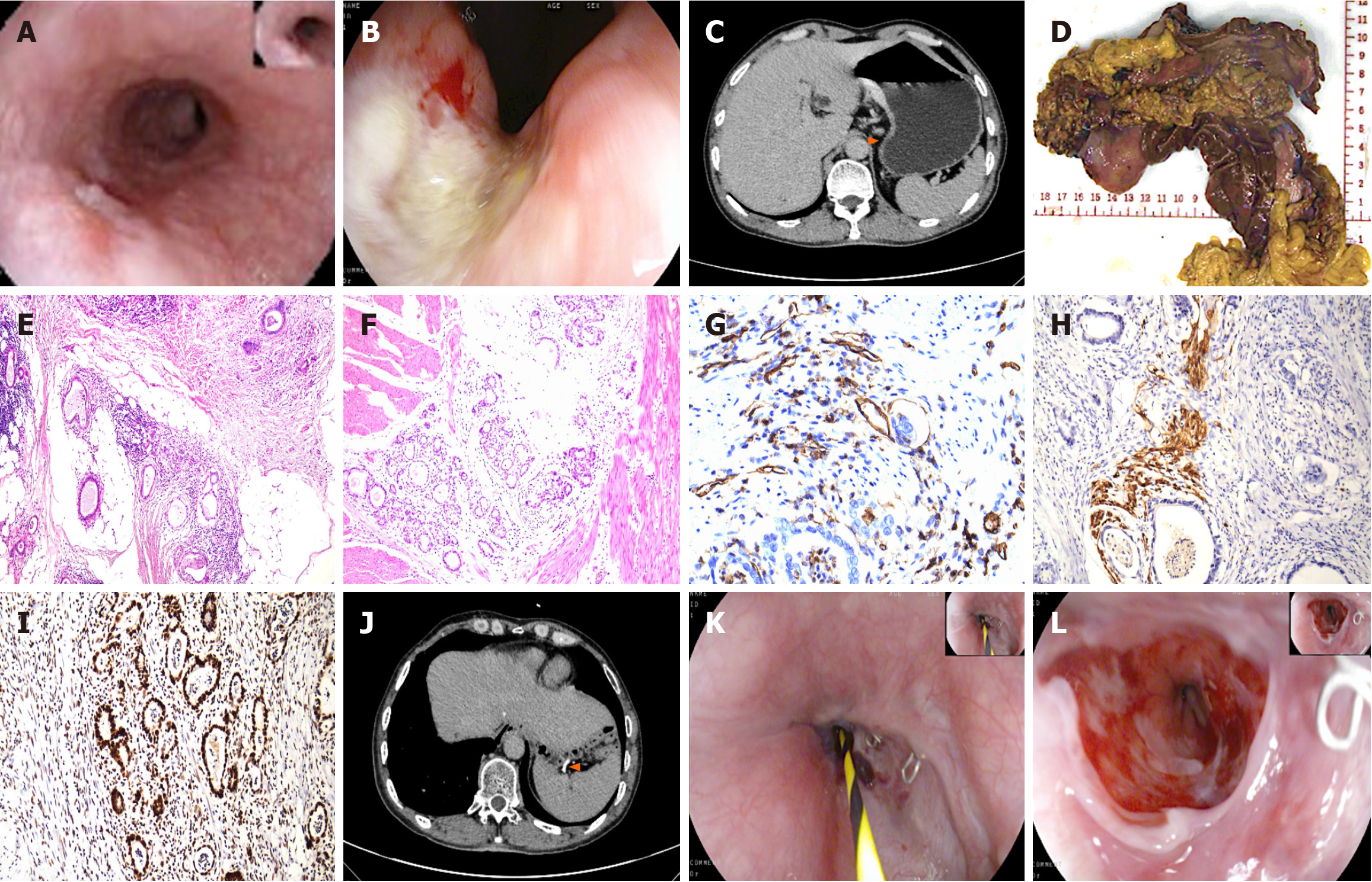
Figure 3 Main examination picture results of Case 3.
A and B: Preoperative endoscopy: Esophagus: A 1.5 cm lesion of the superficial elevated type with central depression. Gastric body: A 3 cm × 4 cm mass covered with yellowish-white moss at the base, surrounded by rough nodular elevations; C: Preoperative abdominal computed tomography (CT); D: Intraoperative macroscopic specimens; E and F: Postoperative pathology (HE, × 100); G-I: Immunohistochemistry of CD31, S-100 and MSH6; J: Postoperative follow-up CT; K and L: Postoperative endoscopy: The esophageal mucosa appeared smooth with white scarring near the lower segment of the anastomosis. The surrounding mucosa was congested, and retained anastomotic staples were visible.
- Citation: Bi XR, Zhao SY, Ma YQ, Duan XY, Hu TT, Bi LZ, Cai HY. Multiple primary cancers with gastrointestinal malignant tumors as the first manifestation: Three case reports and review of literature. World J Gastroenterol 2025; 31(8): 100146
- URL: https://www.wjgnet.com/1007-9327/full/v31/i8/100146.htm
- DOI: https://dx.doi.org/10.3748/wjg.v31.i8.100146