
















©The Author(s) 2025.
World J Gastroenterol. Dec 21, 2025; 31(47): 113205
Published online Dec 21, 2025. doi: 10.3748/wjg.v31.i47.113205
Published online Dec 21, 2025. doi: 10.3748/wjg.v31.i47.113205
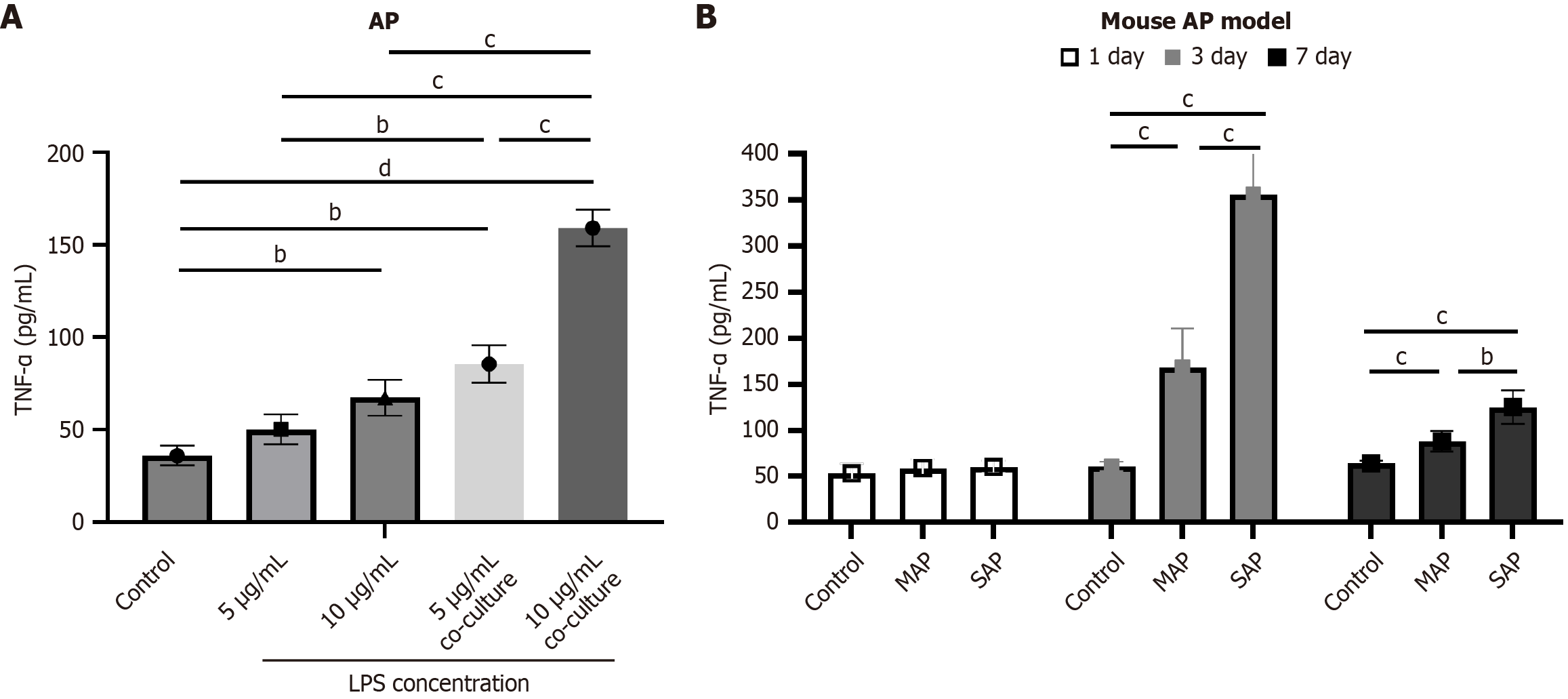
Figure 2 Tumor necrosis factor-α levels were elevated in acute pancreatitis models.
A: Tumor necrosis factor-α levels in conditioned media from MIN-6 cells were quantified by enzyme-linked immunosorbent assay under control conditions, lipopolysaccharide stimulation (5 μg/mL or 10 μg/mL), and co-culture with media from lipopolysaccharide-treated 266-6 cells; B: Serum tumor necrosis factor-α levels in acute pancreatitis mouse models were quantified using the same methodology. Representative results from three independent replicates are shown. bP < 0.01, cP < 0.001, dP < 0.0001. Data are presented as the mean ± SD. AP: Acute pancreatitis; TNF-α: Tumor necrosis factor-α; LPS: Lipopolysaccharide; MAP: Mild acute pancreatitis; SAP: Severe acute pancreatitis.
- Citation: Chen HF, Gong C, Wang TT, Zhu JX, Li WP. Tumor necrosis factor-α promotes abnormal glucose metabolism after acute pancreatitis by inducing islet β-cell apoptosis via Bax/Bcl-2/caspase-3 signaling pathway. World J Gastroenterol 2025; 31(47): 113205
- URL: https://www.wjgnet.com/1007-9327/full/v31/i47/113205.htm
- DOI: https://dx.doi.org/10.3748/wjg.v31.i47.113205