
















©The Author(s) 2025.
World J Gastroenterol. Dec 21, 2025; 31(47): 112705
Published online Dec 21, 2025. doi: 10.3748/wjg.v31.i47.112705
Published online Dec 21, 2025. doi: 10.3748/wjg.v31.i47.112705
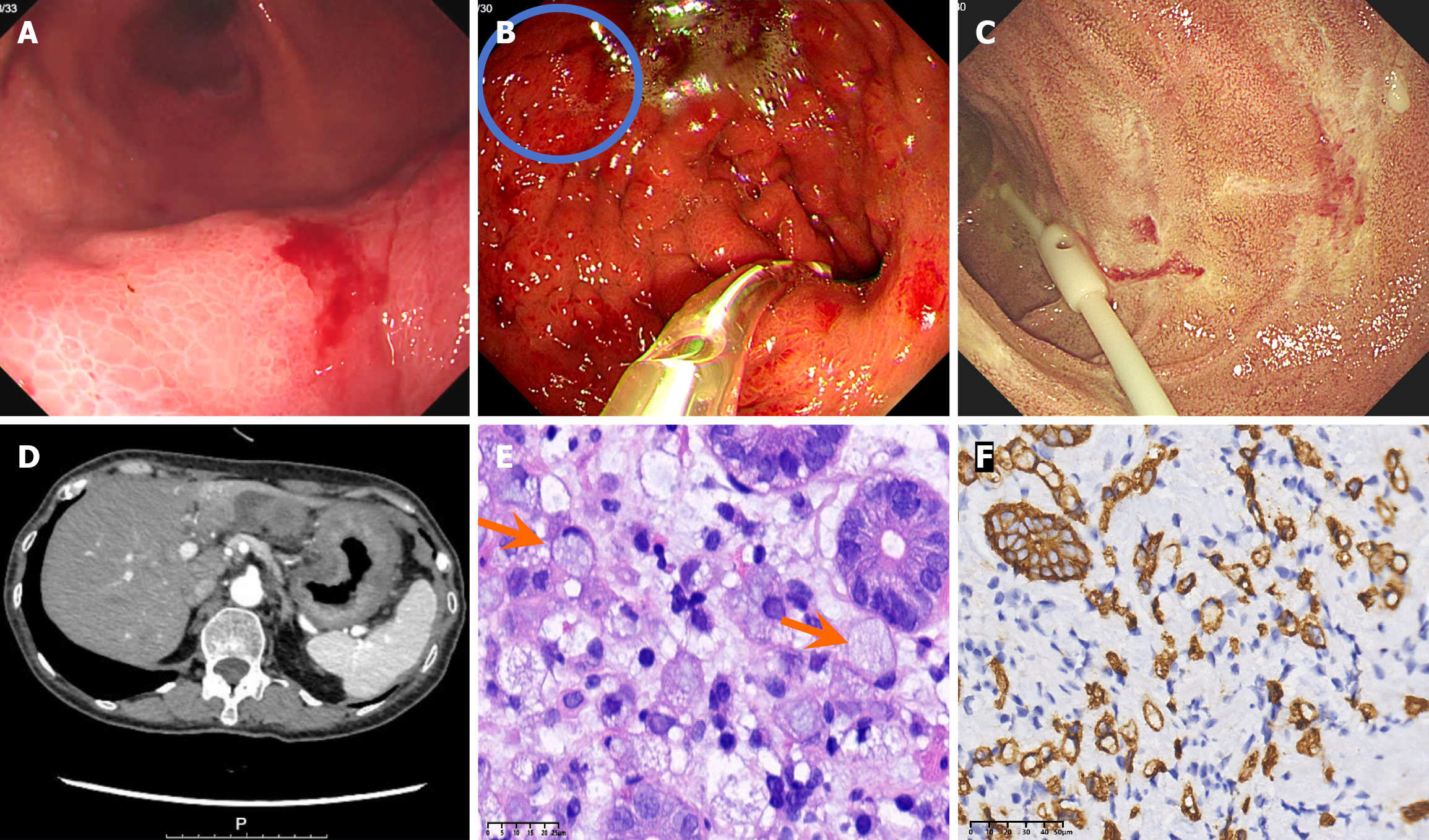
Figure 1 Endoscopic images and computed tomography image of the upper abdomen and pathological characteristics of the lesser curvature of the gastric body.
A: Upper gastrointestinal endoscopy demonstrated diffuse mucosal edema and erythema throughout the gastric fundus, body, and antrum with poor expansion of the gastric cavity after inflation; B: A 1.5 cm mucosal protrusion on the greater curvature; C: Multiple superficial erosions in the descending duodenum with surrounding mucosa exhibiting a finely granular pattern; D: Computed tomography revealed irregular circumferential wall thickening involving the gastric body and antrum and moderate enhancement on an enhanced scan; E: Diffuse infiltrate of the signet-ring cell carcinoma in the lesser curvature of the gastric body (hematoxylin eosin, 400 ×); F: Immunohistochemical staining confirmed that the tumor cells were strongly diffusely positive for CK8 (EnVision method, 200 ×).
- Citation: Chen X, Bo JQ, Gao XX, Zhang SX, Li J, Wang H, Yang MY, Guo QQ, Xiu B, Zeng Y. Indolent T-cell lymphoma of the gastrointestinal tract coexisting with gastric signet-ring cell carcinoma: A case report and review of literature. World J Gastroenterol 2025; 31(47): 112705
- URL: https://www.wjgnet.com/1007-9327/full/v31/i47/112705.htm
- DOI: https://dx.doi.org/10.3748/wjg.v31.i47.112705