
















©The Author(s) 2025.
World J Gastroenterol. Dec 14, 2025; 31(46): 113585
Published online Dec 14, 2025. doi: 10.3748/wjg.v31.i46.113585
Published online Dec 14, 2025. doi: 10.3748/wjg.v31.i46.113585
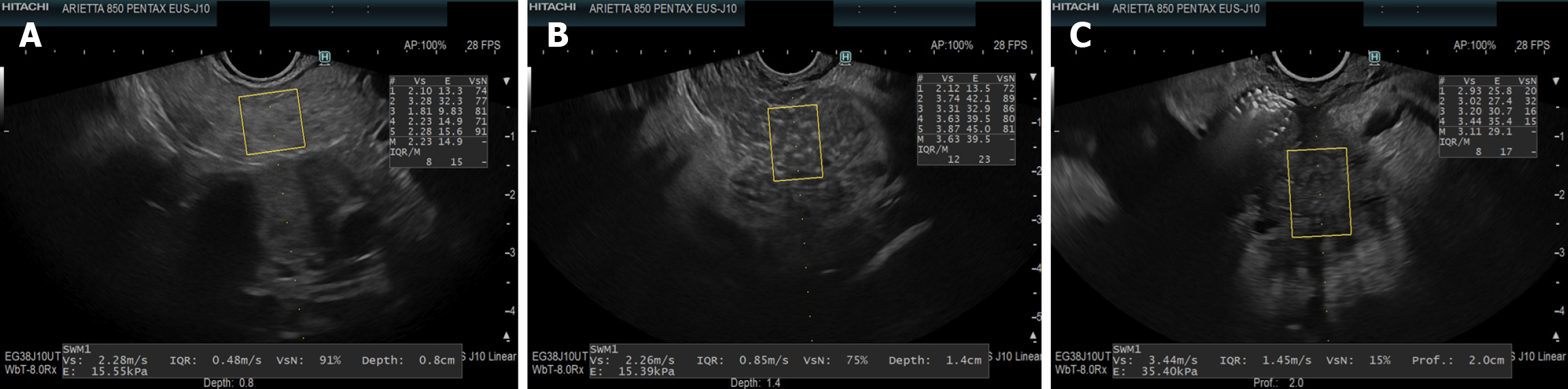
Figure 1 Endoscopic ultrasound-guided shear wave elastography images.
A: Normal pancreas; B: Chronic pancreatitis; C: Pancreatic cancer. In endoscopic ultrasound-guided shear wave elastography, shear wave velocity values were similar in normal parenchyma and chronic pancreatitis, but markedly higher in pancreatic cancer. The net effective shear wave velocity shows a decreasing trend across these conditions, reflecting increasing tissue heterogeneity and stiffness.
- Citation: Paratore M, Miliani S, D’Acunzo G, Viceconti N, Andaloro S, Cerniglia G, Mancuso F, Melita E, Rizzatti G, Gasbarrini A, Riccardi L, Garcovich M. Advances in endoscopic ultrasound-guided shear wave elastography: A comprehensive review of its clinical applications. World J Gastroenterol 2025; 31(46): 113585
- URL: https://www.wjgnet.com/1007-9327/full/v31/i46/113585.htm
- DOI: https://dx.doi.org/10.3748/wjg.v31.i46.113585