
















©The Author(s) 2025.
World J Gastroenterol. Dec 14, 2025; 31(46): 112010
Published online Dec 14, 2025. doi: 10.3748/wjg.v31.i46.112010
Published online Dec 14, 2025. doi: 10.3748/wjg.v31.i46.112010
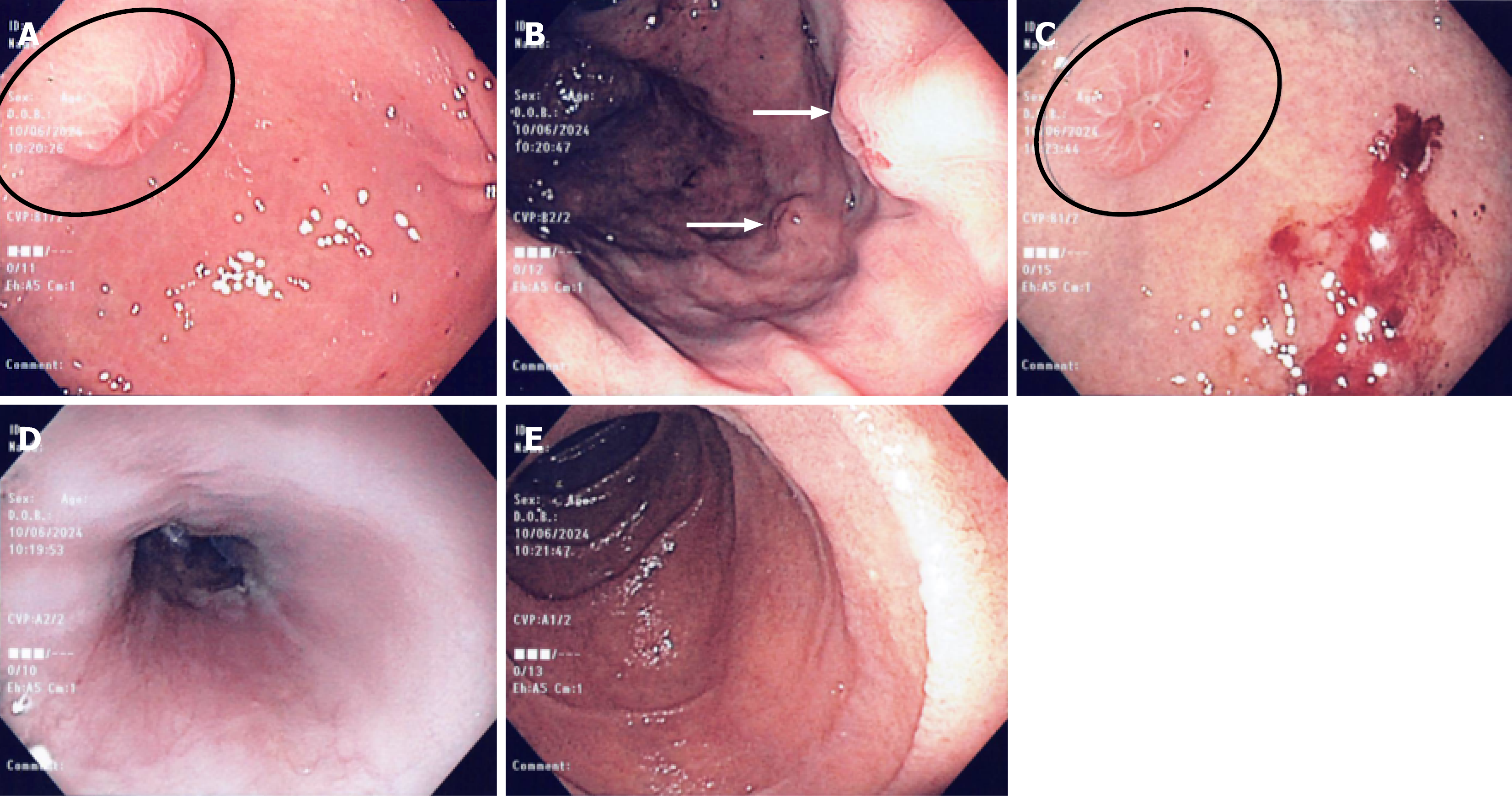
Figure 2 Gastroscopy.
A: Gastroscopy revealing well defined subepithelial nodule in the gastric antrum with a central umbilicated depression (black circle); B: Two subepithelial nodules (white arrows) observed on retroflexion in the gastric body; C: Central umbilication seen in the circled nodule (black circle). Post biopsy heme evident on the adjacent nodule to the right; D: Normal esophagus; E: Normal duodenum.
- Citation: Ahmadi S, Joarder I, Jowhari F. Beyond the skin - metastatic basal cell carcinoma in the stomach: A case report. World J Gastroenterol 2025; 31(46): 112010
- URL: https://www.wjgnet.com/1007-9327/full/v31/i46/112010.htm
- DOI: https://dx.doi.org/10.3748/wjg.v31.i46.112010