
















©The Author(s) 2025.
World J Gastroenterol. Dec 7, 2025; 31(45): 113690
Published online Dec 7, 2025. doi: 10.3748/wjg.v31.i45.113690
Published online Dec 7, 2025. doi: 10.3748/wjg.v31.i45.113690
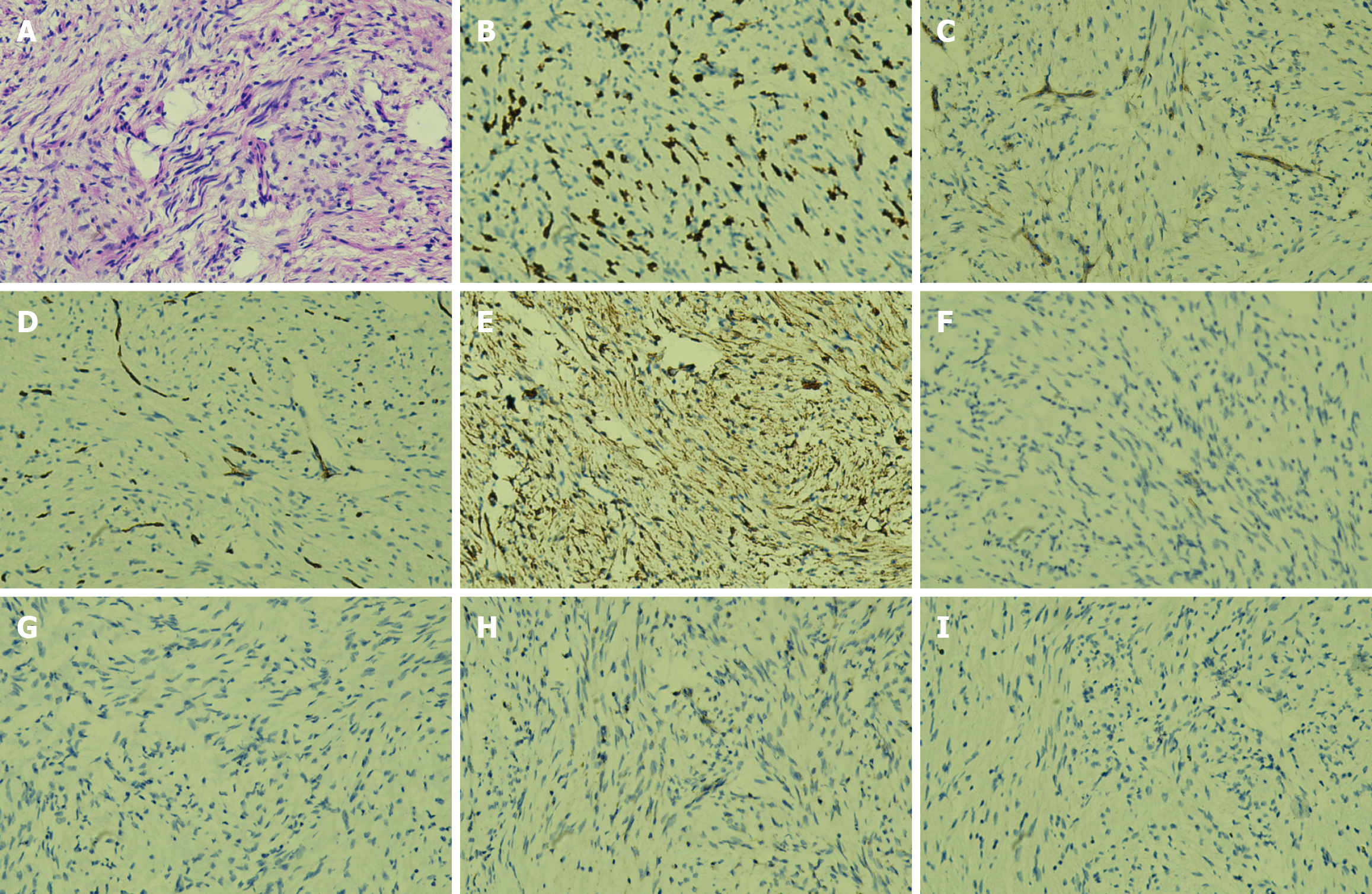
Figure 2 Pathological examination of the tumor.
Pathological findings: The gallbladder measured 9 cm × 3.5 cm × 2.5 cm, with a roughened mucosal surface and mucosal thickness of approximately 0.1 cm. The gallbladder wall measured 0.2 cm, and a localized thickened area measuring 1.8 cm × 1.5 cm × 1.0 cm was identified at the fundus. One gallstone was present, measuring 0.3-0.4 cm in diameter. A: Microscopic examination: Hematoxylin and eosin staining revealed proliferative spindle cells arranged in fascicular and whorled patterns; B-G: Immunohistochemistry: Tumor cells were CD117(+; B), focally positive for DOG-1 (C) and CD34 (D), negative for desmin (F) and S-100 (G), and showed focal positivity for smooth muscle actin (E); H: Succinate dehydrogenase subunit B was negative; I: The Ki-67 proliferation index was approximately 1%. Final diagnosis: Based on the HE morphology and immunohistochemical profile, the lesion was diagnosed as an extra-gastrointestinal stromal tumor. The maximum tumor diameter was approximately 0.4 cm, spindle cell type, with no evident mitotic figures, no significant nuclear atypia, and no necrosis. Risk stratification indicated a very low risk category.
- Citation: Wang P, Zhu CR, Yao J, Xu P, Zhang K, Zhu J, Ge XY, Chen Y, Wang WZ. Primary extra-gastrointestinal stromal tumor of the gallbladder: A case report. World J Gastroenterol 2025; 31(45): 113690
- URL: https://www.wjgnet.com/1007-9327/full/v31/i45/113690.htm
- DOI: https://dx.doi.org/10.3748/wjg.v31.i45.113690