
















©The Author(s) 2025.
World J Gastroenterol. Dec 7, 2025; 31(45): 113690
Published online Dec 7, 2025. doi: 10.3748/wjg.v31.i45.113690
Published online Dec 7, 2025. doi: 10.3748/wjg.v31.i45.113690
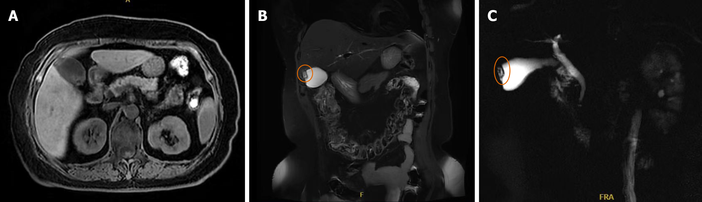
Figure 1 Magnetic resonance cholangiopancreatography.
A-C: A patchy lesion at the gallbladder fundus demonstrated iso-intensity on T1-weighted images and slightly increased signal on T2-weighted images, with small cystic T2 hyperintense foci within, suggesting the possibility of gallbladder adenomyomatosis. The liver, spleen, and pancreas showed no obvious morphological or size abnormalities. No significant abdominal lymphadenopathy or ascites was detected.
- Citation: Wang P, Zhu CR, Yao J, Xu P, Zhang K, Zhu J, Ge XY, Chen Y, Wang WZ. Primary extra-gastrointestinal stromal tumor of the gallbladder: A case report. World J Gastroenterol 2025; 31(45): 113690
- URL: https://www.wjgnet.com/1007-9327/full/v31/i45/113690.htm
- DOI: https://dx.doi.org/10.3748/wjg.v31.i45.113690