
















©The Author(s) 2025.
World J Gastroenterol. Dec 7, 2025; 31(45): 112518
Published online Dec 7, 2025. doi: 10.3748/wjg.v31.i45.112518
Published online Dec 7, 2025. doi: 10.3748/wjg.v31.i45.112518
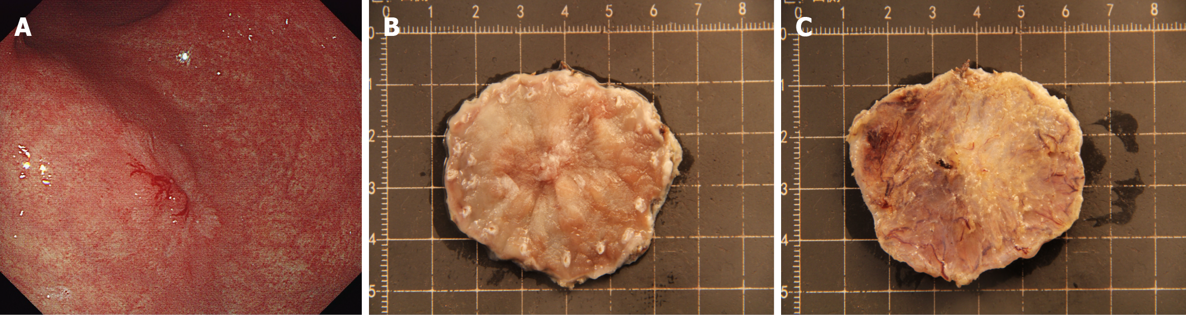
Figure 4 Misdiagnosed case 1 stereomicroscopic overestimation (stereomicroscopy-submucosal vs pathology-T1a).
A: White light imaging of the lesion; B: Stereomicroscopic overview of the formalin-fixed specimen (mucosal surface); C: Stereomicroscopic view of the specimen’s vertical (basal) margin, the central part of the lesion shows localized, non-transparent submucosal tissue with whitish discoloration (diameter > 5 mm), leading to an erroneous diagnosis of submucosal invasion (stereomicroscopy-submucosal). Final histopathology confirmed the lesion was confined to the mucosa (pT1a).
- Citation: Wang J, Chang L, Niu DF, Yan Y, Cao CQ, Li SJ, Wu Q. Diagnostic accuracy of stereomicroscopy assessment of invasion depth in ex vivo specimens of early gastric cancer. World J Gastroenterol 2025; 31(45): 112518
- URL: https://www.wjgnet.com/1007-9327/full/v31/i45/112518.htm
- DOI: https://dx.doi.org/10.3748/wjg.v31.i45.112518