
















©The Author(s) 2025.
World J Gastroenterol. Dec 7, 2025; 31(45): 112518
Published online Dec 7, 2025. doi: 10.3748/wjg.v31.i45.112518
Published online Dec 7, 2025. doi: 10.3748/wjg.v31.i45.112518
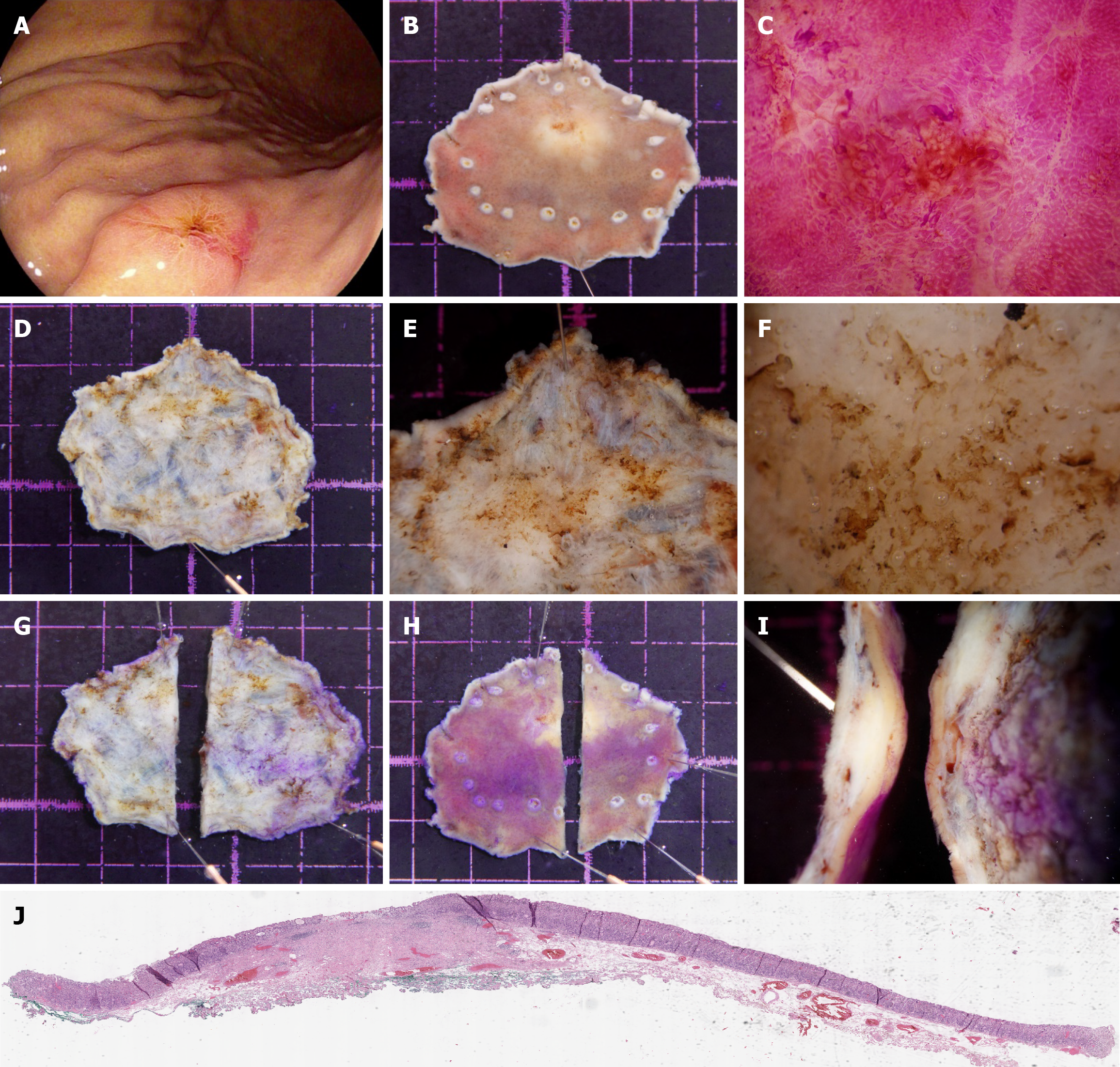
Figure 3 Workflow for preoperative assessment and postoperative management of stereomicroscopy-confirmed submucosal lesions pathologically diagnosed as submucosal.
A: White light imaging of a 0-IIa + IIc lesion located on the posterior wall of the proximal gastric body, exhibiting a central depression; B: Stereomicroscopic evaluation of the specimen after 24-hour formalin fixation; C: High-magnification stereomicroscopy of the central depressed area after eosin staining, demonstrating irregular microsurface patterns; D: Stereomicroscopic overview of the basal surface of the specimen; E: Medium-magnification stereomicroscopy of the basal surface, revealing a thickened submucosal layer with a yellowish-white discoloration; F: High-magnification stereomicroscopy of the basal surface, showing loss of submucosal connective tissue architecture, indicative of tumor invasion into the submucosa; G: The initial incision of the specimen was performed at a distance from the suspected submucosal invasion site on the basal surface to avoid disrupting the area of interest; H: The specimen was inverted to expose the mucosal surface, followed by systematic sampling at 2-mm to 3-mm intervals; I: Lateral view highlighting the deepest point of submucosal invasion; J: Histopathological images confirming the submucosal diagnosis and margin status.
- Citation: Wang J, Chang L, Niu DF, Yan Y, Cao CQ, Li SJ, Wu Q. Diagnostic accuracy of stereomicroscopy assessment of invasion depth in ex vivo specimens of early gastric cancer. World J Gastroenterol 2025; 31(45): 112518
- URL: https://www.wjgnet.com/1007-9327/full/v31/i45/112518.htm
- DOI: https://dx.doi.org/10.3748/wjg.v31.i45.112518