
















©The Author(s) 2025.
World J Gastroenterol. Dec 7, 2025; 31(45): 112518
Published online Dec 7, 2025. doi: 10.3748/wjg.v31.i45.112518
Published online Dec 7, 2025. doi: 10.3748/wjg.v31.i45.112518
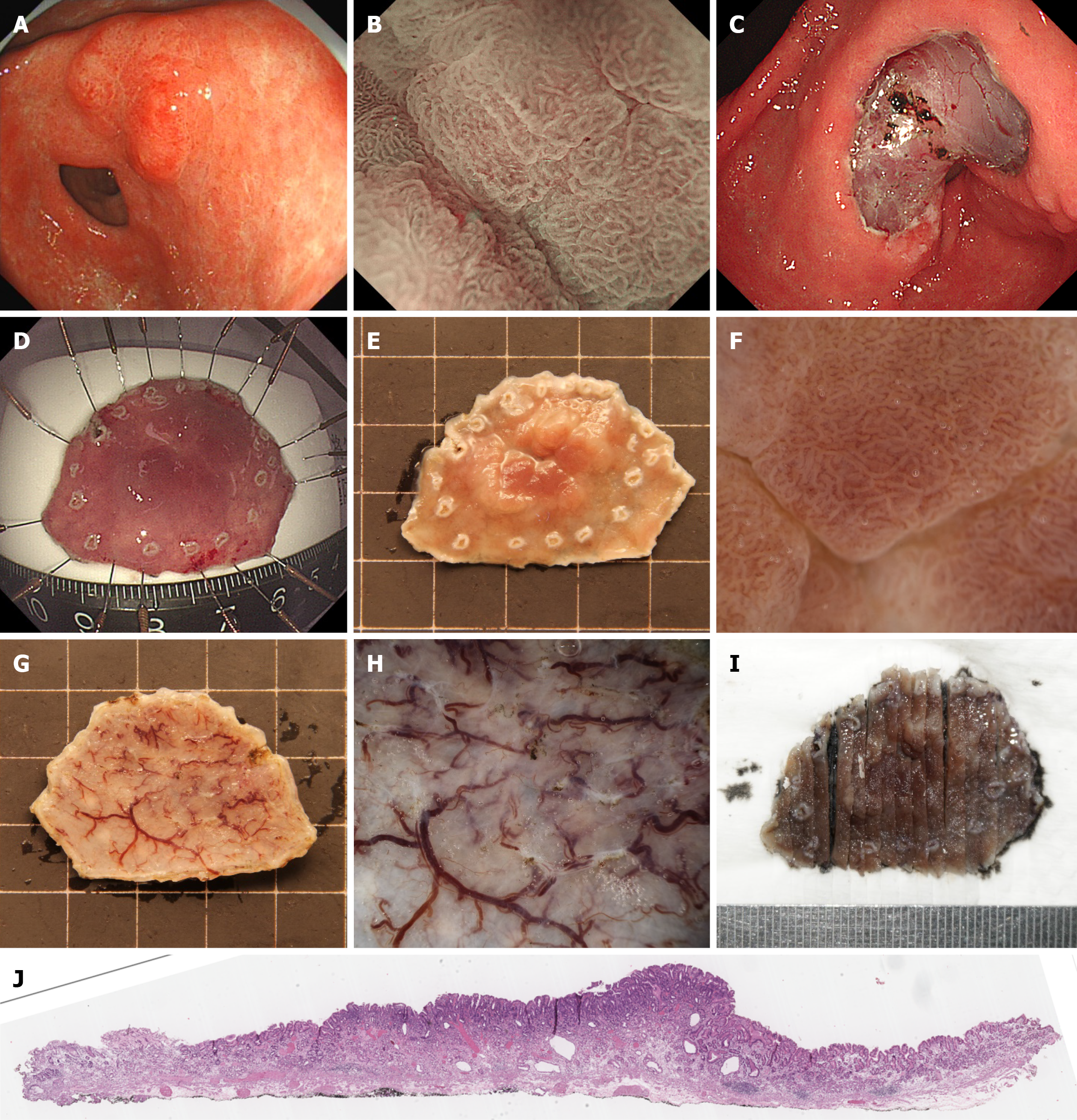
Figure 2 Workflow for preoperative assessment and postoperative management of stereomicroscopy-confirmed mucosal lesions pathologically diagnosed as mucosal.
A: White light imaging of a 0-IIa + IIc lesion in the gastric antrum; B: Magnifying endoscopy with narrow-band imaging of the lesion, demonstrating irregular microsurface and microvascular patterns; C: Post-endoscopic submucosal dissection mucosal defect with visible submucosal vasculature; D: The endoscopic submucosal dissection specimen was spread and pinned on a fixation board for formalin fixation; E: Stereomicroscopic evaluation of the specimen after 24-hour formalin fixation; F: High-magnification stereomicroscopy of the lesion surface, correlating with magnifying endoscopy with narrow-band imaging findings in B, showing irregular microsurface and microvascular architecture; G: Basal surface of the resected specimen under stereomicroscopy; H: High-magnification stereomicroscopy of the basal surface, revealing intact and homogeneous submucosal connective tissue architecture; I: Systematic vertical sectioning of the specimen at 2- to 3-mm intervals, ensuring accurate histopathologic evaluation of margins; J: Histopathological images confirming mucosal (M) diagnosis and margin status.
- Citation: Wang J, Chang L, Niu DF, Yan Y, Cao CQ, Li SJ, Wu Q. Diagnostic accuracy of stereomicroscopy assessment of invasion depth in ex vivo specimens of early gastric cancer. World J Gastroenterol 2025; 31(45): 112518
- URL: https://www.wjgnet.com/1007-9327/full/v31/i45/112518.htm
- DOI: https://dx.doi.org/10.3748/wjg.v31.i45.112518